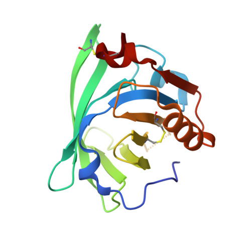
4GNY

Bovine beta-lactoglobulin complex with dodecyl sulfate


Experimental Data Snapshot

  • Method: X-RAY DIFFRACTION
  • Resolution: 1.64 Å
  • R-Value Free: 0.226 
  • R-Value Work: 0.199 
  • R-Value Observed: 0.200 

Starting Model: experimental
View more details

wwPDB Validation   3D Report Full Report


Ligand Structure Quality Assessment 


This is version 1.2 of the entry. See complete history


Literature

Ligand-binding and self-association cooperativity of beta-lactoglobulin

Gutierrez-Magdaleno, G.Bello, M.Portillo-Tellez, M.C.Rodriguez-Romero, A.Garcia-Hernandez, E.

(2013) J Mol Recognit 26: 67-75

  • DOI: https://doi.org/10.1002/jmr.2249
  • Primary Citation of Related Structures:  
    4GNY

  • PubMed Abstract: 

    Unlike most small globular proteins, lipocalins lack a compact hydrophobic core. Instead, they present a large central cavity that functions as the primary binding site for hydrophobic molecules. Not surprisingly, these proteins typically exhibit complex structural dynamics in solution, which is intricately modified by intermolecular recognition events. Although many lipocalins are monomeric, an increasing number of them have been proven to form oligomers. The coupling effects between self-association and ligand binding in these proteins are largely unknown. To address this issue, we have calorimetrically characterized the recognition of dodecyl sulfate by bovine β-lactoglobulin, which forms weak homodimers at neutral pH. A thermodynamic analysis based on coupled-equilibria revealed that dimerization exerts disparate effects on the ligand-binding capacity of β-lactoglobulin. Protein dimerization decreases ligand affinity (or, reciprocally, ligand binding promotes dimer dissociation). The two subunits in the dimer exhibit a positive, entropically driven cooperativity. To investigate the structural determinants of the interaction, the crystal structure of β-lactoglobulin bound to dodecyl sulfate was solved at 1.64 Å resolution.


  • Organizational Affiliation

    Instituto de Química Universidad Nacional Autónoma de México, Circuito Exterior, Ciudad Universitaria, México, DF 04630, México.


Macromolecules
Find similar proteins by:  (by identity cutoff)  |  3D Structure
Entity ID: 1
MoleculeChains Sequence LengthOrganismDetailsImage
Beta-lactoglobulin162Bos taurusMutation(s): 0 
UniProt
Find proteins for P02754 (Bos taurus)
Explore P02754 
Go to UniProtKB:  P02754
Entity Groups  
Sequence Clusters30% Identity50% Identity70% Identity90% Identity95% Identity100% Identity
UniProt GroupP02754
Sequence Annotations
Expand
  • Reference Sequence
Small Molecules
Binding Affinity Annotations 
IDSourceBinding Affinity
SDS PDBBind:  4GNY Kd: 5.20e+4 (nM) from 1 assay(s)
Experimental Data & Validation

Experimental Data

  • Method: X-RAY DIFFRACTION
  • Resolution: 1.64 Å
  • R-Value Free: 0.226 
  • R-Value Work: 0.199 
  • R-Value Observed: 0.200 
  • Space Group: P 32 2 1
Unit Cell:
Length ( Å )Angle ( ˚ )
a = 53.977α = 90
b = 53.977β = 90
c = 112.861γ = 120
Software Package:
Software NamePurpose
CrystalCleardata collection
PHASERphasing
PHENIXrefinement
XDSdata reduction
SCALAdata scaling

Structure Validation

View Full Validation Report


Ligand Structure Quality Assessment 


Entry History 

Deposition Data

Revision History  (Full details and data files)

  • Version 1.0: 2013-02-13
    Type: Initial release
  • Version 1.1: 2023-09-13
    Changes: Data collection, Database references, Derived calculations, Refinement description
  • Version 1.2: 2024-11-06
    Changes: Structure summary