4HAB

Crystal structure of Plk1 Polo-box domain in complex with PL-49


Experimental Data Snapshot

  • Method: X-RAY DIFFRACTION
  • Resolution: 2.65 Å
  • R-Value Free: 
    0.259 (Depositor), 0.300 (DCC) 
  • R-Value Work: 
    0.260 (Depositor), 0.250 (DCC) 
  • R-Value Observed: 
    0.260 (Depositor) 

wwPDB Validation   3D Report Full Report


Ligand Structure Quality Assessment 


This is version 1.3 of the entry. See complete history


Literature

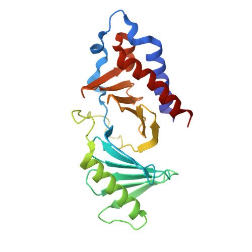
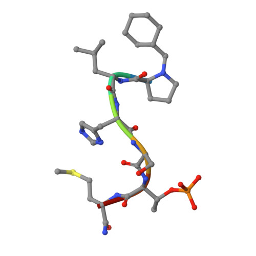
Development of cyclic peptomer inhibitors targeting the polo-box domain of polo-like kinase 1.

Murugan, R.N.Park, J.E.Lim, D.Ahn, M.Cheong, C.Kwon, T.Nam, K.Y.Choi, S.H.Kim, B.Y.Yoon, D.Y.Yaffe, M.B.Yu, D.Y.Lee, K.S.Bang, J.K.

(2013) Bioorg Med Chem 21: 2623-2634

  • DOI: https://doi.org/10.1016/j.bmc.2013.02.020
  • Primary Citation of Related Structures:  
    4HAB, 4HY2

  • PubMed Abstract: 

    The polo-box domain (PBD) of polo-like kinase 1 (Plk1) is essentially required for the function of Plk1 in cell proliferation. The availability of the phosphopeptide-binding pocket on PBD provides a unique opportunity to develop novel protein-protein interaction inhibitors. Recent identification of a minimal 5-residue-long phosphopeptide, PLHSpT, as a Plk1 PBD-specific ligand has led to the development of several peptide-based inhibitors, but none of them is cyclic peptide. Through the combination of single-peptoid mimics and thio-ether bridged cyclization, we successfully demonstrated for the first time two cyclic peptomers, PL-116 and PL-120, dramatically improved the binding affinity without losing mono-specificity against Plk1 PBD in comparison with the linear parental peptide, PLHSpT. These cyclic peptomers could serve as promising templates for future drug designs to inhibit Plk1 PBD.


  • Organizational Affiliation

    Division of Magnetic Resonance, Korea Basic Science Institute, Ochang, Chung-Buk 363-883, Cheongwon, Republic of Korea.


Macromolecules
Find similar proteins by:  (by identity cutoff)  |  3D Structure
Entity ID: 1
MoleculeChains Sequence LengthOrganismDetailsImage
Serine/threonine-protein kinase PLK1
A, B, C
223Homo sapiensMutation(s): 0 
Gene Names: PLKPLK1
EC: 2.7.11.21
UniProt & NIH Common Fund Data Resources
Find proteins for P53350 (Homo sapiens)
Explore P53350 
Go to UniProtKB:  P53350
PHAROS:  P53350
GTEx:  ENSG00000166851 
Entity Groups  
Sequence Clusters30% Identity50% Identity70% Identity90% Identity95% Identity100% Identity
UniProt GroupP53350
Sequence Annotations
Expand
  • Reference Sequence

Find similar proteins by:  Sequence   |   3D Structure  

Entity ID: 2
MoleculeChains Sequence LengthOrganismDetailsImage
PL-49
D, E, F
7synthetic constructMutation(s): 0 
Sequence Annotations
Expand
  • Reference Sequence
Biologically Interesting Molecules (External Reference) 1 Unique
Experimental Data & Validation

Experimental Data

  • Method: X-RAY DIFFRACTION
  • Resolution: 2.65 Å
  • R-Value Free:  0.259 (Depositor), 0.300 (DCC) 
  • R-Value Work:  0.260 (Depositor), 0.250 (DCC) 
  • R-Value Observed: 0.260 (Depositor) 
Space Group: P 43 21 2
Unit Cell:
Length ( Å )Angle ( ˚ )
a = 95.043α = 90
b = 95.043β = 90
c = 240.58γ = 90
Software Package:
Software NamePurpose
SCALAdata scaling
MOLREPphasing
CNSrefinement
PDB_EXTRACTdata extraction
SERGUIdata collection

Structure Validation

View Full Validation Report


Ligand Structure Quality Assessment 


Entry History 

Deposition Data

Revision History  (Full details and data files)

  • Version 1.0: 2013-04-03
    Type: Initial release
  • Version 1.1: 2013-05-22
    Changes: Database references
  • Version 1.2: 2017-11-15
    Changes: Refinement description, Source and taxonomy
  • Version 1.3: 2022-12-21
    Changes: Database references, Derived calculations