4HJJ

Structure Reveals Function of the Dual Variable Domain Immunoglobulin (DVD-Ig) Molecule


Experimental Data Snapshot

  • Method: X-RAY DIFFRACTION
  • Resolution: 2.10 Å
  • R-Value Free: 0.220 
  • R-Value Work: 0.184 
  • R-Value Observed: 0.186 

wwPDB Validation   3D Report Full Report


This is version 1.0 of the entry. See complete history


Literature

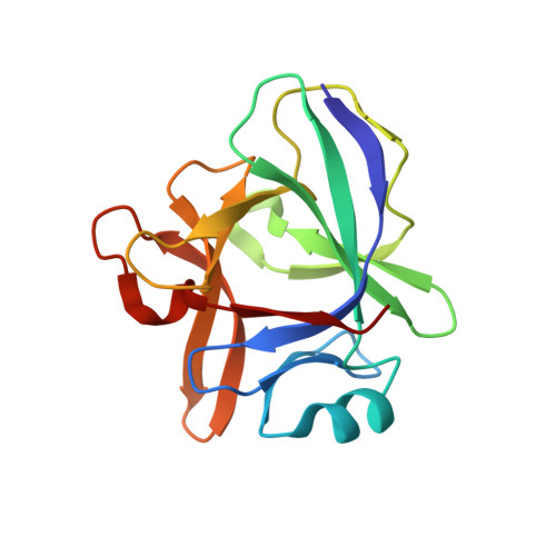
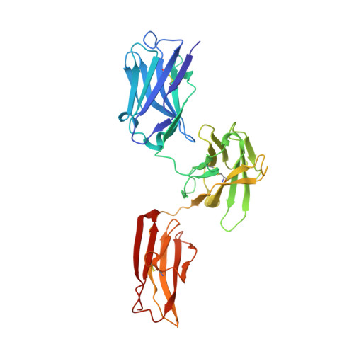
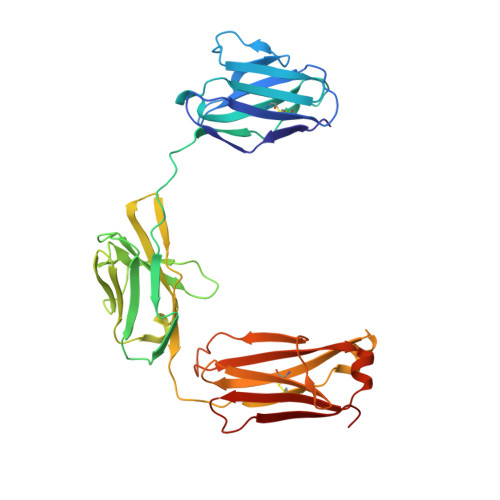
Structure reveals function of the dual variable domain immunoglobulin (DVD-Ig[TM]) molecule

Jakob, C.G.Edalji, R.Judge, R.A.Digiammarino, E.Li, Y.Gu, J.Ghayur, T.

(2013) MAbs 5: 358-363

  • DOI: https://doi.org/10.4161/mabs.23977
  • Primary Citation of Related Structures:  
    4HJJ

  • PubMed Abstract: 

    Several bispecific antibody-based formats have been developed over the past 25 years in an effort to produce a new generation of immunotherapeutics that target two or more disease mechanisms simultaneously. One such format, the dual-variable domain immunoglobulin (DVD-Ig™), combines the target binding domains of two monoclonal antibodies via flexible naturally occurring linkers, which yields a tetravalent IgG - like molecule. We report the structure of an interleukin (IL)12-IL18 DVD-Ig™ Fab (DFab) fragment with IL18 bound to the inner variable domain (VD) that reveals the remarkable flexibility of the DVD-Ig™ molecule and how the DVD-Ig™ format can function to bind four antigens simultaneously. An understanding of how the inner variable domain retains function is of critical importance for designing DVD-Ig™ molecules, and for better understanding of the flexibility of immunoglobulin variable domains and linkers, which may aid in the design of improved bi- and multi-specific biologics in general.


  • Organizational Affiliation

    Department of Structural Biology; AbbVie Inc.; North Chicago, IL USA.


Macromolecules
Find similar proteins by:  (by identity cutoff)  |  3D Structure
Entity ID: 1
MoleculeChains Sequence LengthOrganismDetailsImage
Interleukin-18157Homo sapiensMutation(s): 4 
Gene Names: IL18IGIFIL1F4
UniProt & NIH Common Fund Data Resources
Find proteins for Q14116 (Homo sapiens)
Explore Q14116 
Go to UniProtKB:  Q14116
PHAROS:  Q14116
GTEx:  ENSG00000150782 
Entity Groups  
Sequence Clusters30% Identity50% Identity70% Identity90% Identity95% Identity100% Identity
UniProt GroupQ14116
Sequence Annotations
Expand
  • Reference Sequence
Find similar proteins by:  (by identity cutoff)  |  3D Structure
Entity ID: 2
MoleculeChains Sequence LengthOrganismDetailsImage
Anti-IL12 Anti-IL18 DFab Heavy ChainB [auth H]348Homo sapiensMutation(s): 0 
Entity Groups  
Sequence Clusters30% Identity50% Identity70% Identity90% Identity95% Identity100% Identity
Sequence Annotations
Expand
  • Reference Sequence
Find similar proteins by:  (by identity cutoff)  |  3D Structure
Entity ID: 3
MoleculeChains Sequence LengthOrganismDetailsImage
Anti-IL12 Anti-IL18 DFab Light ChainC [auth L]327Homo sapiensMutation(s): 0 
Entity Groups  
Sequence Clusters30% Identity50% Identity70% Identity90% Identity95% Identity100% Identity
Sequence Annotations
Expand
  • Reference Sequence
Experimental Data & Validation

Experimental Data

  • Method: X-RAY DIFFRACTION
  • Resolution: 2.10 Å
  • R-Value Free: 0.220 
  • R-Value Work: 0.184 
  • R-Value Observed: 0.186 
  • Space Group: C 1 2 1
Unit Cell:
Length ( Å )Angle ( ˚ )
a = 274.385α = 90
b = 65.498β = 95.2
c = 78.802γ = 90
Software Package:
Software NamePurpose
BUSTER-TNTrefinement
PDB_EXTRACTdata extraction
ADSCdata collection
HKL-2000data reduction
HKL-2000data scaling
PHASERphasing
MOLREPphasing
BUSTERrefinement

Structure Validation

View Full Validation Report


Entry History 

Deposition Data

Revision History  (Full details and data files)

  • Version 1.0: 2013-05-08
    Type: Initial release