4I1B

FUNCTIONAL IMPLICATIONS OF INTERLEUKIN-1BETA BASED ON THE THREE-DIMENSIONAL STRUCTURE


Experimental Data Snapshot

  • Method: X-RAY DIFFRACTION
  • Resolution: 2.00 Å
  • R-Value Observed: 0.190 

wwPDB Validation   3D Report Full Report


This is version 1.3 of the entry. See complete history


Literature

Functional implications of interleukin-1 beta based on the three-dimensional structure.

Veerapandian, B.Gilliland, G.L.Raag, R.Svensson, A.L.Masui, Y.Hirai, Y.Poulos, T.L.

(1992) Proteins 12: 10-23

  • DOI: https://doi.org/10.1002/prot.340120103
  • Primary Citation of Related Structures:  
    4I1B

  • PubMed Abstract: 

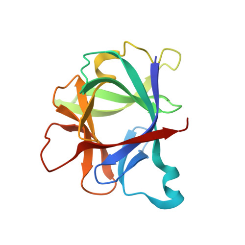
    The molecular structure of interleukin-1 beta, a hormone-like cytokine with roles in several disease processes, has been determined at 2.0 A resolution and refined to a crystallographic R-factor of 0.19. The framework of this molecule consists of 12 antiparallel beta-strands exhibiting pseudo-3-fold symmetry. Six of the strands make up a beta-barrel with polar residues concentrated at either end. Analysis of the three-dimensional structure, together with results from site-directed mutagenesis and biochemical and immunological studies, suggest that the core of the beta-barrel plays an important functional role. A large patch of charged residues on one end of the barrel is proposed as the binding surface with which IL-1 interacts with its receptor.


  • Organizational Affiliation

    Center for Advanced Research in Biotechnology, University of Maryland, Rockville.


Macromolecules
Find similar proteins by:  (by identity cutoff)  |  3D Structure
Entity ID: 1
MoleculeChains Sequence LengthOrganismDetailsImage
INTERLEUKIN-1 BETA153Homo sapiensMutation(s): 0 
UniProt & NIH Common Fund Data Resources
Find proteins for P01584 (Homo sapiens)
Explore P01584 
Go to UniProtKB:  P01584
PHAROS:  P01584
GTEx:  ENSG00000125538 
Entity Groups  
Sequence Clusters30% Identity50% Identity70% Identity90% Identity95% Identity100% Identity
UniProt GroupP01584
Sequence Annotations
Expand
  • Reference Sequence
Experimental Data & Validation

Experimental Data

  • Method: X-RAY DIFFRACTION
  • Resolution: 2.00 Å
  • R-Value Observed: 0.190 
  • Space Group: P 43
Unit Cell:
Length ( Å )Angle ( ˚ )
a = 55.14α = 90
b = 55.14β = 90
c = 76.66γ = 90
Software Package:
Software NamePurpose
RESTRAINrefinement

Structure Validation

View Full Validation Report


Entry History 

Deposition Data

Revision History  (Full details and data files)

  • Version 1.0: 1990-04-15
    Type: Initial release
  • Version 1.1: 2008-03-25
    Changes: Version format compliance
  • Version 1.2: 2011-07-13
    Changes: Version format compliance
  • Version 1.3: 2024-02-28
    Changes: Data collection, Database references, Other