4I1Q | pdb_00004i1q

Crystal Structure of hBRAP1 N-BAR domain


Experimental Data Snapshot

  • Method: X-RAY DIFFRACTION
  • Resolution: 2.53 Å
  • R-Value Free: 
    0.252 (Depositor), 0.251 (DCC) 
  • R-Value Work: 
    0.196 (Depositor), 0.195 (DCC) 
  • R-Value Observed: 
    0.199 (Depositor) 

wwPDB Validation 3D Report Full Report

Validation slider image for 4I1Q

This is version 1.1 of the entry. See complete history

Literature

Bin2 Is a Membrane Sculpting N-BAR Protein That Influences Leucocyte Podosomes, Motility and Phagocytosis

Sanchez-Barrena, M.J.Vallis, Y.Clatworthy, M.R.Doherty, G.J.Veprintsev, D.B.Evans, P.R.McMahon, H.T.

(2012) PLoS One 7: e52401-e52401

  • DOI: https://doi.org/10.1371/journal.pone.0052401
  • Primary Citation Related Structures: 
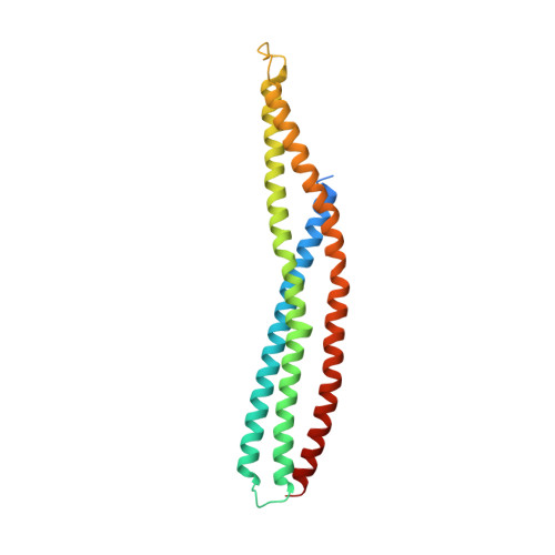
    4I1Q

  • PubMed Abstract: 

    Cell motility, adhesion and phagocytosis are controlled by actin and membrane remodelling processes. Bridging integrator-2 (Bin2) also called Breast cancer-associated protein 1 (BRAP1) is a predicted N-BAR domain containing protein with unknown function that is highly expressed in leucocytic cells. In the present study we solved the structure of Bin2 BAR domain and studied its membrane binding and bending properties in vitro and in vivo. Live-cell imaging experiments showed that Bin2 is associated with actin rich structures on the plasma membrane, where it was targeted through its N-BAR domain. Pull-down experiments and immunoprecipitations showed that Bin2 C-terminus bound SH3 domain containing proteins such as Endophilin A2 and α-PIX. siRNA of endogenous protein led to decreased cell migration, increased phagocytosis and reduced podosome density and dynamics. In contrast, overexpression of Bin2 led to decreased phagocytosis and increased podosome density and dynamics. We conclude that Bin2 is a membrane-sculpting protein that influences podosome formation, motility and phagocytosis in leucocytes. Further understanding of this protein may be key to understand the behaviour of leucocytes under physiological and pathological conditions.


  • Organizational Affiliation
    • MRC Laboratory of Molecular Biology, Cambridge, United Kingdom. xmjose@iqfr.csic.es

Macromolecule Content 

  • Total Structure Weight: 52.13 kDa 
  • Atom Count: 3,393 
  • Modeled Residue Count: 398 
  • Deposited Residue Count: 442 
  • Unique protein chains: 1

Macromolecules

Find similar proteins by:|  3D Structure
Entity ID: 1
MoleculeChains  Sequence LengthOrganismDetailsImage
Bridging integrator 2
A, B
221Homo sapiensMutation(s): 0 
Gene Names: BIN2
UniProt & NIH Common Fund Data Resources
Find proteins for Q9UBW5 (Homo sapiens)
Explore Q9UBW5 
Go to UniProtKB:  Q9UBW5
PHAROS:  Q9UBW5
GTEx:  ENSG00000110934 
Entity Groups
Sequence Clusters30% Identity50% Identity70% Identity90% Identity95% Identity100% Identity
UniProt GroupQ9UBW5
Sequence Annotations
Expand
Reference Sequence

Experimental Data & Validation

Experimental Data

  • Method: X-RAY DIFFRACTION
  • Resolution: 2.53 Å
  • R-Value Free:  0.252 (Depositor), 0.251 (DCC) 
  • R-Value Work:  0.196 (Depositor), 0.195 (DCC) 
  • R-Value Observed: 0.199 (Depositor) 
Space Group: P 21 21 21
Unit Cell:
Length ( Å )Angle ( ˚ )
a = 79.147α = 90
b = 81.278β = 90
c = 81.101γ = 90
Software Package:
Software NamePurpose
DNAdata collection
SHARPphasing
REFMACrefinement
MOSFLMdata reduction
SCALAdata scaling

Structure Validation

View Full Validation Report


Entry History 

Deposition Data

Revision History  (Full details and data files)

  • Version 1.0: 2013-02-06
    Type: Initial release
  • Version 1.1: 2024-03-20
    Changes: Data collection, Database references, Refinement description