4I7Y

Crystal Structure of Human Alpha Thrombin in Complex with a 27-mer Aptamer Bound to Exosite II


Experimental Data Snapshot

  • Method: X-RAY DIFFRACTION
  • Resolution: 2.40 Å
  • R-Value Free: 
    0.249 (Depositor), 0.249 (DCC) 
  • R-Value Work: 
    0.181 (Depositor), 0.180 (DCC) 
  • R-Value Observed: 
    0.184 (Depositor) 

wwPDB Validation   3D Report Full Report


Ligand Structure Quality Assessment 

Created with Raphaël 2.3.0Worse 01 BetterLigand structure goodness of fit to experimental dataBest fitted 0G6Click on this verticalbar to view detailsBest fitted NAGClick on this verticalbar to view details

This is version 1.4 of the entry. See complete history


Literature

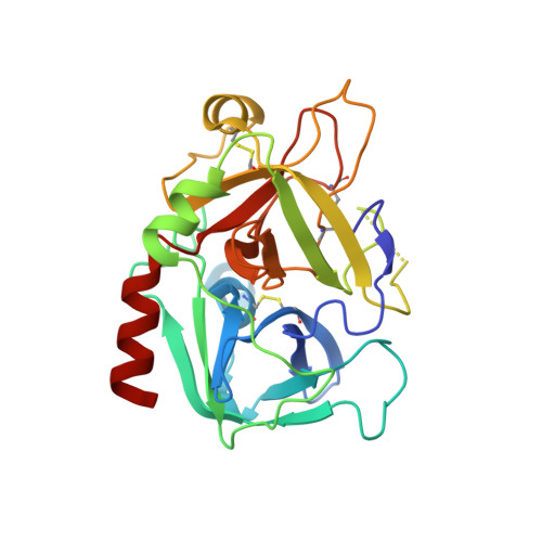
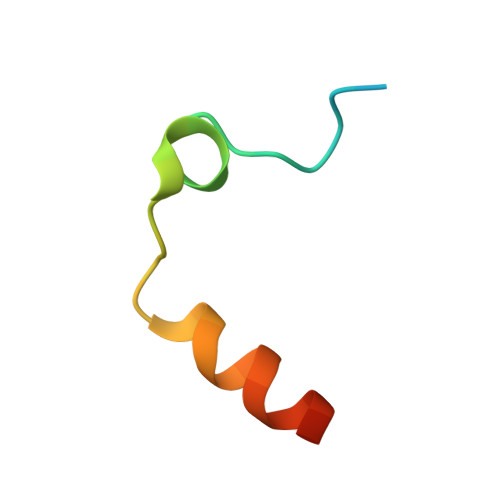
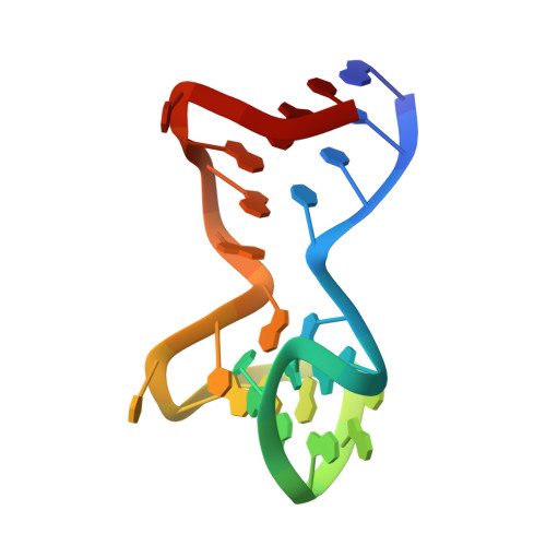
Duplex-quadruplex motifs in a peculiar structural organization cooperatively contribute to thrombin binding of a DNA aptamer.

Russo Krauss, I.Pica, A.Merlino, A.Mazzarella, L.Sica, F.

(2013) Acta Crystallogr D Biol Crystallogr 69: 2403-2411

  • DOI: https://doi.org/10.1107/S0907444913022269
  • Primary Citation of Related Structures:  
    4I7Y

  • PubMed Abstract: 

    Potent second-generation thrombin aptamers adopt a duplex-quadruplex bimodular folding and recognize thrombin exosite II with very high affinity and specificity. A sound model of these oligonucleotides, either free or in complex with thrombin, is not yet available. Here, a structural study of one of these aptamers, HD22-27mer, is presented. The crystal structure of this aptamer in complex with thrombin displays a novel architecture in which the helical stem is enchained to a pseudo-G-quadruplex. The results also underline the role of the residues that join the duplex and quadruplex motifs and control their recruitment in thrombin binding.


  • Organizational Affiliation

    Department of Chemical Sciences, University of Naples `Federico II', Complesso Universitario di Monte Sant'Angelo, I-80126 Naples, Italy.


Macromolecules

Find similar proteins by:  (by identity cutoff)  |  3D Structure
Entity ID: 1
MoleculeChains Sequence LengthOrganismDetailsImage
ProthrombinA [auth H]259Homo sapiensMutation(s): 0 
EC: 3.4.21.5
UniProt & NIH Common Fund Data Resources
Find proteins for P00734 (Homo sapiens)
Explore P00734 
Go to UniProtKB:  P00734
PHAROS:  P00734
GTEx:  ENSG00000180210 
Entity Groups  
Sequence Clusters30% Identity50% Identity70% Identity90% Identity95% Identity100% Identity
UniProt GroupP00734
Glycosylation
Glycosylation Sites: 1Go to GlyGen: P00734-1
Sequence Annotations
Expand
  • Reference Sequence
Find similar proteins by:  (by identity cutoff)  |  3D Structure
Entity ID: 2
MoleculeChains Sequence LengthOrganismDetailsImage
ProthrombinB [auth L]36Homo sapiensMutation(s): 0 
EC: 3.4.21.5
UniProt & NIH Common Fund Data Resources
Find proteins for P00734 (Homo sapiens)
Explore P00734 
Go to UniProtKB:  P00734
PHAROS:  P00734
GTEx:  ENSG00000180210 
Entity Groups  
Sequence Clusters30% Identity50% Identity70% Identity90% Identity95% Identity100% Identity
UniProt GroupP00734
Sequence Annotations
Expand
  • Reference Sequence
Small Molecules
Ligands 3 Unique
IDChains Name / Formula / InChI Key2D Diagram3D Interactions
0G6
Query on 0G6

Download Ideal Coordinates CCD File 
D [auth H]D-phenylalanyl-N-[(2S,3S)-6-{[amino(iminio)methyl]amino}-1-chloro-2-hydroxyhexan-3-yl]-L-prolinamide
C21 H34 Cl N6 O3
DVFLYEYCMMLBTQ-VSZNYVQBSA-O
NAG
Query on NAG

Download Ideal Coordinates CCD File 
F [auth L]2-acetamido-2-deoxy-beta-D-glucopyranose
C8 H15 N O6
OVRNDRQMDRJTHS-FMDGEEDCSA-N
NA
Query on NA

Download Ideal Coordinates CCD File 
E [auth H]SODIUM ION
Na
FKNQFGJONOIPTF-UHFFFAOYSA-N
Biologically Interesting Molecules (External Reference) 1 Unique
Experimental Data & Validation

Experimental Data

  • Method: X-RAY DIFFRACTION
  • Resolution: 2.40 Å
  • R-Value Free:  0.249 (Depositor), 0.249 (DCC) 
  • R-Value Work:  0.181 (Depositor), 0.180 (DCC) 
  • R-Value Observed: 0.184 (Depositor) 
Space Group: P 1 21 1
Unit Cell:
Length ( Å )Angle ( ˚ )
a = 48.144α = 90
b = 81.904β = 99.66
c = 53.72γ = 90
Software Package:
Software NamePurpose
DENZOdata reduction
PHASERphasing
REFMACrefinement
HKL-2000data reduction
HKL-2000data scaling

Structure Validation

View Full Validation Report


Ligand Structure Quality Assessment 

Created with Raphaël 2.3.0Worse 01 BetterLigand structure goodness of fit to experimental dataBest fitted 0G6Click on this verticalbar to view detailsBest fitted NAGClick on this verticalbar to view details

Entry History 

Deposition Data

Revision History  (Full details and data files)

  • Version 1.0: 2013-10-16
    Type: Initial release
  • Version 1.1: 2013-12-11
    Changes: Database references
  • Version 1.2: 2014-01-01
    Changes: Database references
  • Version 1.3: 2020-07-29
    Type: Remediation
    Reason: Carbohydrate remediation
    Changes: Data collection, Derived calculations, Structure summary
  • Version 1.4: 2024-10-09
    Changes: Data collection, Database references, Structure summary