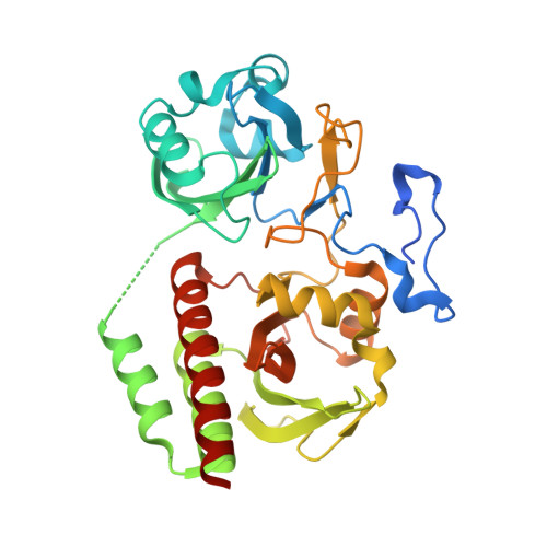
4IJG

Crystal structure of monomeric bacteriophytochrome


Experimental Data Snapshot

  • Method: X-RAY DIFFRACTION
  • Resolution: 1.70 Å
  • R-Value Free: 0.227 
  • R-Value Work: 0.205 
  • R-Value Observed: 0.206 

Starting Model: experimental
View more details

wwPDB Validation   3D Report Full Report


Ligand Structure Quality Assessment 


This is version 1.4 of the entry. See complete history


Literature

Origins of fluorescence in evolved bacteriophytochromes.

Bhattacharya, S.Auldridge, M.E.Lehtivuori, H.Ihalainen, J.A.Forest, K.T.

(2014) J Biol Chem 289: 32144-32152

  • DOI: https://doi.org/10.1074/jbc.M114.589739
  • Primary Citation of Related Structures:  
    4IJG, 4O8G

  • PubMed Abstract: 

    Use of fluorescent proteins to study in vivo processes in mammals requires near-infrared (NIR) biomarkers that exploit the ability of light in this range to penetrate tissue. Bacteriophytochromes (BphPs) are photoreceptors that couple absorbance of NIR light to photoisomerization, protein conformational changes, and signal transduction. BphPs have been engineered to form NIR fluorophores, including IFP1.4, Wi-Phy, and the iRFP series, initially by replacement of Asp-207 by His. This position was suggestive because its main chain carbonyl is within hydrogen-bonding distance to pyrrole ring nitrogens of the biliverdin chromophore, thus potentially functioning as a crucial transient proton sink during photoconversion. To explain the origin of fluorescence in these phytofluors, we solved the crystal structures of IFP1.4 and a comparison non-fluorescent monomeric phytochrome DrCBDmon. Met-186 and Val-288 in IFP1.4 are responsible for the formation of a tightly packed hydrophobic hub around the biliverdin D ring. Met-186 is also largely responsible for the blue-shifted IFP1.4 excitation maximum relative to the parent BphP. The structure of IFP1.4 revealed decreased structural heterogeneity and a contraction of two surface regions as direct consequences of side chain substitutions. Unexpectedly, IFP1.4 with Asp-207 reinstalled (IFPrev) has a higher fluorescence quantum yield (∼9%) than most NIR phytofluors published to date. In agreement, fluorescence lifetime measurements confirm the exceptionally long excited state lifetimes, up to 815 ps, in IFP1.4 and IFPrev. Our research helps delineate the origin of fluorescence in engineered BphPs and will facilitate the wide-spread adoption of phytofluors as biomarkers.


  • Organizational Affiliation

    Departments of Bacteriology, University of Wisconsin, Madison, Wisconsin 53706 and.


Macromolecules
Find similar proteins by:  (by identity cutoff)  |  3D Structure
Entity ID: 1
MoleculeChains Sequence LengthOrganismDetailsImage
Bacteriophytochrome341Deinococcus radiodurans R1 = ATCC 13939 = DSM 20539Mutation(s): 0 
Gene Names: bphPDR_A0050
EC: 2.7.13.3
UniProt
Find proteins for Q9RZA4 (Deinococcus radiodurans (strain ATCC 13939 / DSM 20539 / JCM 16871 / CCUG 27074 / LMG 4051 / NBRC 15346 / NCIMB 9279 / VKM B-1422 / R1))
Explore Q9RZA4 
Go to UniProtKB:  Q9RZA4
Entity Groups  
Sequence Clusters30% Identity50% Identity70% Identity90% Identity95% Identity100% Identity
UniProt GroupQ9RZA4
Sequence Annotations
Expand
  • Reference Sequence
Small Molecules
Ligands 4 Unique
IDChains Name / Formula / InChI Key2D Diagram3D Interactions
LBV
Query on LBV

Download Ideal Coordinates CCD File 
B [auth A]3-[2-[(Z)-[3-(2-carboxyethyl)-5-[(Z)-(4-ethenyl-3-methyl-5-oxidanylidene-pyrrol-2-ylidene)methyl]-4-methyl-pyrrol-1-ium -2-ylidene]methyl]-5-[(Z)-[(3E)-3-ethylidene-4-methyl-5-oxidanylidene-pyrrolidin-2-ylidene]methyl]-4-methyl-1H-pyrrol-3- yl]propanoic acid
C33 H37 N4 O6
DKMLMZVDTGOEGU-ISEYCTJISA-O
PEG
Query on PEG

Download Ideal Coordinates CCD File 
F [auth A]DI(HYDROXYETHYL)ETHER
C4 H10 O3
MTHSVFCYNBDYFN-UHFFFAOYSA-N
PO4
Query on PO4

Download Ideal Coordinates CCD File 
E [auth A]PHOSPHATE ION
O4 P
NBIIXXVUZAFLBC-UHFFFAOYSA-K
GOL
Query on GOL

Download Ideal Coordinates CCD File 
C [auth A],
D [auth A]
GLYCEROL
C3 H8 O3
PEDCQBHIVMGVHV-UHFFFAOYSA-N
Experimental Data & Validation

Experimental Data

  • Method: X-RAY DIFFRACTION
  • Resolution: 1.70 Å
  • R-Value Free: 0.227 
  • R-Value Work: 0.205 
  • R-Value Observed: 0.206 
  • Space Group: C 1 2 1
Unit Cell:
Length ( Å )Angle ( ˚ )
a = 94.563α = 90
b = 54.816β = 91.8
c = 69.77γ = 90
Software Package:
Software NamePurpose
SCALEPACKdata scaling
PHASERphasing
REFMACrefinement
PDB_EXTRACTdata extraction
HKL-2000data reduction

Structure Validation

View Full Validation Report


Ligand Structure Quality Assessment 


Entry History 

Deposition Data

Revision History  (Full details and data files)

  • Version 1.0: 2013-12-25
    Type: Initial release
  • Version 1.1: 2014-10-01
    Changes: Database references
  • Version 1.2: 2014-10-08
    Changes: Database references
  • Version 1.3: 2014-12-03
    Changes: Database references
  • Version 1.4: 2023-09-20
    Changes: Data collection, Database references, Derived calculations, Refinement description, Structure summary