4ILW

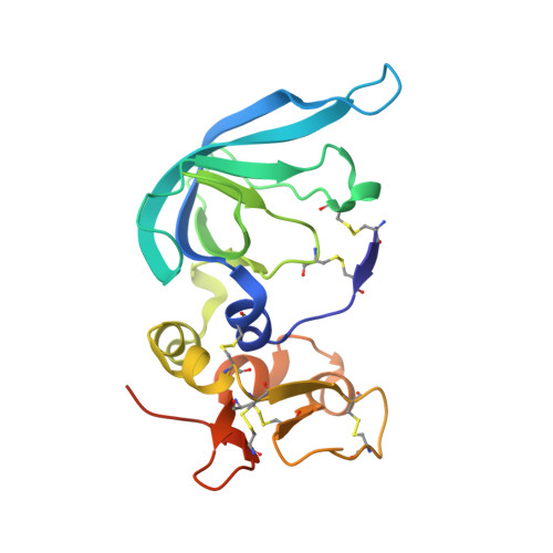
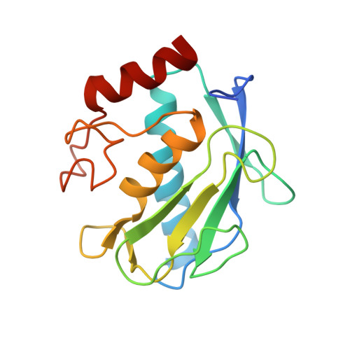
Complex of matrix metalloproteinase-10 catalytic domain (MMP-10cd) with tissue inhibitor of metalloproteinases-2 (TIMP-2)


Experimental Data Snapshot

  • Method: X-RAY DIFFRACTION
  • Resolution: 2.10 Å
  • R-Value Free: 0.263 
  • R-Value Work: 0.216 
  • R-Value Observed: 0.219 

Starting Models: experimental
View more details

wwPDB Validation   3D Report Full Report


This is version 1.2 of the entry. See complete history


Literature

Matrix Metalloproteinase-10/TIMP-2 Structure and Analyses Define Conserved Core Interactions and Diverse Exosite Interactions in MMP/TIMP Complexes.

Batra, J.Soares, A.S.Mehner, C.Radisky, E.S.

(2013) PLoS One 8: e75836-e75836

  • DOI: https://doi.org/10.1371/journal.pone.0075836
  • Primary Citation of Related Structures:  
    4ILW

  • PubMed Abstract: 

    Matrix metalloproteinases (MMPs) play central roles in vertebrate tissue development, remodeling, and repair. The endogenous tissue inhibitors of metalloproteinases (TIMPs) regulate proteolytic activity by binding tightly to the MMP active site. While each of the four TIMPs can inhibit most MMPs, binding data reveal tremendous heterogeneity in affinities of different TIMP/MMP pairs, and the structural features that differentiate stronger from weaker complexes are poorly understood. Here we report the crystal structure of the comparatively weakly bound human MMP-10/TIMP-2 complex at 2.1 Å resolution. Comparison with previously reported structures of MMP-3/TIMP-1, MT1-MMP/TIMP-2, MMP-13/TIMP-2, and MMP-10/TIMP-1 complexes offers insights into the structural basis of binding selectivity. Our analyses identify a group of highly conserved contacts at the heart of MMP/TIMP complexes that define the conserved mechanism of inhibition, as well as a second category of diverse adventitious contacts at the periphery of the interfaces. The AB loop of the TIMP N-terminal domain and the contact loops of the TIMP C-terminal domain form highly variable peripheral contacts that can be considered as separate exosite interactions. In some complexes these exosite contacts are extensive, while in other complexes the AB loop or C-terminal domain contacts are greatly reduced and appear to contribute little to complex stability. Our data suggest that exosite interactions can enhance MMP/TIMP binding, although in the relatively weakly bound MMP-10/TIMP-2 complex they are not well optimized to do so. Formation of highly variable exosite interactions may provide a general mechanism by which TIMPs are fine-tuned for distinct regulatory roles in biology.


  • Organizational Affiliation

    Department of Cancer Biology, Mayo Clinic Cancer Center, Jacksonville, Florida, United States of America.


Macromolecules
Find similar proteins by:  (by identity cutoff)  |  3D Structure
Entity ID: 1
MoleculeChains Sequence LengthOrganismDetailsImage
Metalloproteinase inhibitor 2
A, B
194Homo sapiensMutation(s): 0 
Gene Names: TIMP2
UniProt & NIH Common Fund Data Resources
Find proteins for P16035 (Homo sapiens)
Explore P16035 
Go to UniProtKB:  P16035
PHAROS:  P16035
GTEx:  ENSG00000035862 
Entity Groups  
Sequence Clusters30% Identity50% Identity70% Identity90% Identity95% Identity100% Identity
UniProt GroupP16035
Sequence Annotations
Expand
  • Reference Sequence
Find similar proteins by:  (by identity cutoff)  |  3D Structure
Entity ID: 2
MoleculeChains Sequence LengthOrganismDetailsImage
Stromelysin-2C [auth D],
D [auth F]
165Homo sapiensMutation(s): 0 
Gene Names: MMP-10MMP10STMY2
EC: 3.4.24.22
UniProt & NIH Common Fund Data Resources
Find proteins for P09238 (Homo sapiens)
Explore P09238 
Go to UniProtKB:  P09238
PHAROS:  P09238
GTEx:  ENSG00000166670 
Entity Groups  
Sequence Clusters30% Identity50% Identity70% Identity90% Identity95% Identity100% Identity
UniProt GroupP09238
Sequence Annotations
Expand
  • Reference Sequence
Small Molecules
Ligands 2 Unique
IDChains Name / Formula / InChI Key2D Diagram3D Interactions
ZN
Query on ZN

Download Ideal Coordinates CCD File 
E [auth D],
F [auth D],
J [auth F],
K [auth F]
ZINC ION
Zn
PTFCDOFLOPIGGS-UHFFFAOYSA-N
CA
Query on CA

Download Ideal Coordinates CCD File 
G [auth D]
H [auth D]
I [auth D]
L [auth F]
M [auth F]
G [auth D],
H [auth D],
I [auth D],
L [auth F],
M [auth F],
N [auth F]
CALCIUM ION
Ca
BHPQYMZQTOCNFJ-UHFFFAOYSA-N
Experimental Data & Validation

Experimental Data

  • Method: X-RAY DIFFRACTION
  • Resolution: 2.10 Å
  • R-Value Free: 0.263 
  • R-Value Work: 0.216 
  • R-Value Observed: 0.219 
  • Space Group: P 1
Unit Cell:
Length ( Å )Angle ( ˚ )
a = 37.687α = 76.14
b = 56.934β = 79.84
c = 82.592γ = 71.25
Software Package:
Software NamePurpose
HKL-2000data collection
PHASERphasing
REFMACrefinement
DENZOdata reduction
SCALEPACKdata scaling

Structure Validation

View Full Validation Report


Entry History 

Deposition Data

Revision History  (Full details and data files)

  • Version 1.0: 2013-11-13
    Type: Initial release
  • Version 1.1: 2023-09-20
    Changes: Data collection, Database references, Derived calculations, Refinement description
  • Version 1.2: 2024-10-30
    Changes: Structure summary