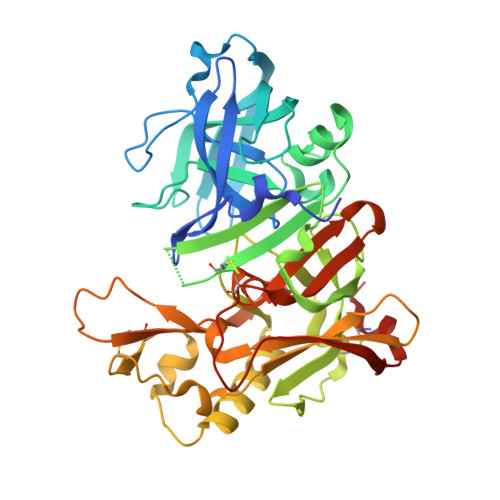
4J0V

CRYSTAL STRUCTURE OF BACE-1 IN COMPLEX WITH 5-Cyano-pyridine-2-carboxylic acid [3-((4R,5R)-2-amino-5-fluoro-4-methyl-5,6-dihydro-4H-[1,3]oxazin-4-yl)-4-fluoro-phenyl]-amide


Experimental Data Snapshot

  • Method: X-RAY DIFFRACTION
  • Resolution: 1.94 Å
  • R-Value Free: 0.207 
  • R-Value Work: 0.168 
  • R-Value Observed: 0.170 

wwPDB Validation   3D Report Full Report


Ligand Structure Quality Assessment 


This is version 1.2 of the entry. See complete history


Literature

beta-Secretase (BACE1) Inhibitors with High In Vivo Efficacy Suitable for Clinical Evaluation in Alzheimer s Disease

Hilpert, H.Guba, W.Woltering, T.J.Wostl, W.Pinard, E.Mauser, H.Mayweg, A.V.Rogers-Evans, M.Humm, R.Krummenacher, D.Muser, T.Schnider, C.Jacobsen, H.Ozmen, L.Bergadano, A.Banner, D.W.Hochstrasser, R.Kuglstatter, A.David-Pierson, P.Fischer, H.Polara, A.Narquizian, R.

(2013) J Med Chem 56: 3980-3995

  • DOI: https://doi.org/10.1021/jm400225m
  • Primary Citation of Related Structures:  
    3ZLQ, 3ZMG, 4J0P, 4J0T, 4J0V, 4J0Y, 4J0Z, 4J17, 4J1C, 4J1E, 4J1F, 4J1H, 4J1I, 4J1K

  • PubMed Abstract: 

    An extensive fluorine scan of 1,3-oxazines revealed the power of fluorine(s) to lower the pKa and thereby dramatically change the pharmacological profile of this class of BACE1 inhibitors. The CF3 substituted oxazine 89, a potent and highly brain penetrant BACE1 inhibitor, was able to reduce significantly CSF Aβ40 and 42 in rats at oral doses as low as 1 mg/kg. The effect was long lasting, showing a significant reduction of Aβ40 and 42 even after 24 h. In contrast to 89, compound 1b lacking the CF3 group was virtually inactive in vivo.


  • Organizational Affiliation

    Discovery Chemistry, Pharma Research & Early Development, Grenzacherstrasse 124, Basel CH-4070, Switzerland. hans.hilpert@roche.com


Macromolecules
Find similar proteins by:  (by identity cutoff)  |  3D Structure
Entity ID: 1
MoleculeChains Sequence LengthOrganismDetailsImage
Beta-secretase 1409Homo sapiensMutation(s): 1 
Gene Names: BACE1BACEKIAA1149
EC: 3.4.23.46
UniProt & NIH Common Fund Data Resources
Find proteins for P56817 (Homo sapiens)
Explore P56817 
Go to UniProtKB:  P56817
PHAROS:  P56817
GTEx:  ENSG00000186318 
Entity Groups  
Sequence Clusters30% Identity50% Identity70% Identity90% Identity95% Identity100% Identity
UniProt GroupP56817
Sequence Annotations
Expand
  • Reference Sequence
Small Molecules
Ligands 3 Unique
IDChains Name / Formula / InChI Key2D Diagram3D Interactions
1H7
Query on 1H7

Download Ideal Coordinates CCD File 
E [auth A]N-{3-[(4R,5R)-2-amino-5-fluoro-4-methyl-5,6-dihydro-4H-1,3-oxazin-4-yl]-4-fluorophenyl}-5-cyanopyridine-2-carboxamide
C18 H15 F2 N5 O2
ALKHFUPBQSAGMS-MAUKXSAKSA-N
DMS
Query on DMS

Download Ideal Coordinates CCD File 
D [auth A]DIMETHYL SULFOXIDE
C2 H6 O S
IAZDPXIOMUYVGZ-UHFFFAOYSA-N
NA
Query on NA

Download Ideal Coordinates CCD File 
B [auth A],
C [auth A]
SODIUM ION
Na
FKNQFGJONOIPTF-UHFFFAOYSA-N
Binding Affinity Annotations 
IDSourceBinding Affinity
1H7 PDBBind:  4J0V IC50: 28 (nM) from 1 assay(s)
Experimental Data & Validation

Experimental Data

  • Method: X-RAY DIFFRACTION
  • Resolution: 1.94 Å
  • R-Value Free: 0.207 
  • R-Value Work: 0.168 
  • R-Value Observed: 0.170 
  • Space Group: P 61 2 2
Unit Cell:
Length ( Å )Angle ( ˚ )
a = 101.928α = 90
b = 101.928β = 90
c = 170.068γ = 120
Software Package:
Software NamePurpose
RemDAqdata collection
REFMACrefinement
XDSdata reduction
SCALAdata scaling
REFMACphasing

Structure Validation

View Full Validation Report


Ligand Structure Quality Assessment 


Entry History 

Deposition Data

Revision History  (Full details and data files)

  • Version 1.0: 2013-05-01
    Type: Initial release
  • Version 1.1: 2013-06-19
    Changes: Database references
  • Version 1.2: 2017-11-15
    Changes: Refinement description