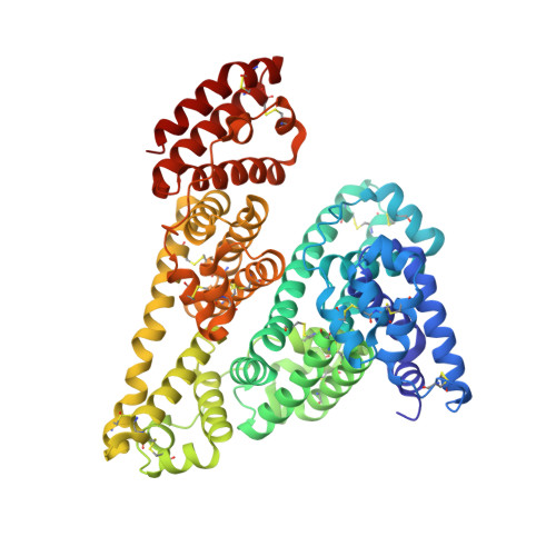
4J2V

Crystal Structure of Equine Serum Albumin in complex with 3,5-diiodosalicylic acid


Experimental Data Snapshot

  • Method: X-RAY DIFFRACTION
  • Resolution: 2.12 Å
  • R-Value Free: 0.238 
  • R-Value Work: 0.181 
  • R-Value Observed: 0.183 

Starting Model: experimental
View more details

wwPDB Validation   3D Report Full Report


Ligand Structure Quality Assessment 


This is version 1.3 of the entry. See complete history


Literature

Crystallographic studies of the complexes of bovine and equine serum albumin with 3,5-diiodosalicylic acid.

Sekula, B.Zielinski, K.Bujacz, A.

(2013) Int J Biol Macromol 60C: 316-324

  • DOI: https://doi.org/10.1016/j.ijbiomac.2013.06.004
  • Primary Citation of Related Structures:  
    4J2V, 4JK4

  • PubMed Abstract: 

    Due to their extraordinary binding properties, serum albumins are the main transporters of many small molecules in the circulatory system. Although all mammalian serum albumins exhibit quite high sequence similarity, their binding abilities are not the same. Until now, only human serum albumin (HSA) was subjected to extensive structural studies in complexes with various ligands. Here we present two crystal structures of the complexes of equine and bovine serum albumins with 3,5-diiodosalicylic acid (DIS), at resolutions 2.12 Å and 2.65 Å, respectively, and analyze interactions of the DIS ligand with both macromolecules. We highlight the differences in distribution of DIS binding sites between the bovine and equine serum albumins and compare results with the HSA binding ability of DIS and other structurally similar ligands.


  • Organizational Affiliation

    Institute of Technical Biochemistry, Lodz University of Technology, Stefanowskiego 4/10, 90-924 Lodz, Poland.


Macromolecules
Find similar proteins by:  (by identity cutoff)  |  3D Structure
Entity ID: 1
MoleculeChains Sequence LengthOrganismDetailsImage
Serum albumin583Equus caballusMutation(s): 0 
UniProt
Find proteins for P35747 (Equus caballus)
Explore P35747 
Go to UniProtKB:  P35747
Entity Groups  
Sequence Clusters30% Identity50% Identity70% Identity90% Identity95% Identity100% Identity
UniProt GroupP35747
Sequence Annotations
Expand
  • Reference Sequence
Small Molecules
Ligands 4 Unique
IDChains Name / Formula / InChI Key2D Diagram3D Interactions
DIU
Query on DIU

Download Ideal Coordinates CCD File 
B [auth A],
C [auth A],
D [auth A],
E [auth A]
2-HYDROXY-3,5-DIIODO-BENZOIC ACID
C7 H4 I2 O3
DHZVWQPHNWDCFS-UHFFFAOYSA-N
MLI
Query on MLI

Download Ideal Coordinates CCD File 
F [auth A]
G [auth A]
H [auth A]
I [auth A]
J [auth A]
F [auth A],
G [auth A],
H [auth A],
I [auth A],
J [auth A],
K [auth A],
L [auth A]
MALONATE ION
C3 H2 O4
OFOBLEOULBTSOW-UHFFFAOYSA-L
ACT
Query on ACT

Download Ideal Coordinates CCD File 
U [auth A]ACETATE ION
C2 H3 O2
QTBSBXVTEAMEQO-UHFFFAOYSA-M
FMT
Query on FMT

Download Ideal Coordinates CCD File 
M [auth A]
N [auth A]
O [auth A]
P [auth A]
Q [auth A]
M [auth A],
N [auth A],
O [auth A],
P [auth A],
Q [auth A],
R [auth A],
S [auth A],
T [auth A]
FORMIC ACID
C H2 O2
BDAGIHXWWSANSR-UHFFFAOYSA-N
Experimental Data & Validation

Experimental Data

  • Method: X-RAY DIFFRACTION
  • Resolution: 2.12 Å
  • R-Value Free: 0.238 
  • R-Value Work: 0.181 
  • R-Value Observed: 0.183 
  • Space Group: P 61
Unit Cell:
Length ( Å )Angle ( ˚ )
a = 88.88α = 90
b = 88.88β = 90
c = 134.34γ = 120
Software Package:
Software NamePurpose
PHENIXrefinement
REFMACrefinement
XDSdata scaling
XDSdata reduction
REFMACphasing

Structure Validation

View Full Validation Report


Ligand Structure Quality Assessment 


Entry History 

Deposition Data

Revision History  (Full details and data files)

  • Version 1.0: 2013-07-24
    Type: Initial release
  • Version 1.1: 2019-07-17
    Changes: Data collection, Refinement description
  • Version 1.2: 2023-09-20
    Changes: Data collection, Database references, Derived calculations, Refinement description
  • Version 1.3: 2024-11-06
    Changes: Structure summary