4JAX | pdb_00004jax

Crystal structure of dimeric KlHxk1 in crystal form X


Experimental Data Snapshot

  • Method: X-RAY DIFFRACTION
  • Resolution: 2.26 Å
  • R-Value Free: 
    0.240 (Depositor), 0.239 (DCC) 
  • R-Value Work: 
    0.200 (Depositor), 0.199 (DCC) 
  • R-Value Observed: 
    0.201 (Depositor) 

Starting Model: experimental
View more details

wwPDB Validation 3D Report Full Report

Validation slider image for 4JAX

This is version 1.4 of the entry. See complete history

Literature

In vivo phosphorylation and in vitro autophosphorylation-inactivation of Kluyveromyces lactis hexokinase KlHxk1.

Kettner, K.Kuettner, E.B.Otto, A.Lilie, H.Golbik, R.P.Strater, N.Kriegel, T.M.

(2013) Biochem Biophys Res Commun 435: 313-318

  • DOI: https://doi.org/10.1016/j.bbrc.2013.03.121
  • Primary Citation Related Structures: 
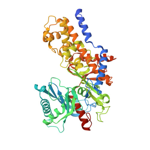
    4JAX

  • PubMed Abstract: 

    The bifunctional hexokinase KlHxk1 is a key component of glucose-dependent signal transduction in Kluyveromyces lactis. KlHxk1 is phosphorylated in vivo and undergoes ATP-dependent autophosphorylation-inactivation in vitro. This study identifies serine-15 as the site of in vivo phosphorylation and serine-157 as the autophosphorylation-inactivation site. X-ray crystallography of the in vivo phosphorylated enzyme indicates the existence of a ring-shaped symmetrical homodimer carrying two phosphoserine-15 residues. In contrast, small-angle X-ray scattering and equilibrium sedimentation analyses reveal the existence of monomeric phosphoserine-15 KlHxk1 in solution. While phosphorylation at serine-15 and concomitant homodimer dissociation are likely to be involved in glucose signalling, mechanism and putative physiological significance of KlHxk1 inactivation by autophosphorylation at serine-157 remain to be established.


  • Organizational Affiliation
    • Institute of Physiological Chemistry, Carl Gustav Carus Medical Faculty, Technische Universität Dresden, Dresden, Germany. karina.kettner@tu-dresden.de

Macromolecule Content 

  • Total Structure Weight: 324.86 kDa 
  • Atom Count: 22,473 
  • Modeled Residue Count: 2,821 
  • Deposited Residue Count: 2,910 
  • Unique protein chains: 1

Macromolecules

Find similar proteins by:|  3D Structure
Entity ID: 1
MoleculeChains  Sequence LengthOrganismDetailsImage
Hexokinase
A, B, C, D, E
A, B, C, D, E, F
485Kluyveromyces lactis NRRL Y-1140Mutation(s): 0 
Gene Names: KLLA0D11352gRAG5
EC: 2.7.1.1
UniProt
Find proteins for P33284 (Kluyveromyces lactis (strain ATCC 8585 / CBS 2359 / DSM 70799 / NBRC 1267 / NRRL Y-1140 / WM37))
Explore P33284 
Go to UniProtKB:  P33284
Entity Groups
Sequence Clusters30% Identity50% Identity70% Identity90% Identity95% Identity100% Identity
UniProt GroupP33284
Sequence Annotations
Expand
Reference Sequence

Small Molecules

Ligands 2 Unique
IDChains Name / Formula / InChI Key2D Diagram3D Interactions
PO4

Query on PO4


Download:Ideal Coordinates CCD File
AA [auth E]
BA [auth E]
EA [auth F]
G [auth A]
H [auth A]
AA [auth E],
BA [auth E],
EA [auth F],
G [auth A],
H [auth A],
I [auth A],
J [auth A],
M [auth B],
O [auth C],
P [auth C],
Q [auth C],
R [auth C],
U [auth D],
V [auth D],
W [auth D],
X [auth D],
Z [auth E]
PHOSPHATE ION
O4 P
NBIIXXVUZAFLBC-UHFFFAOYSA-K
GOL

Query on GOL


Download:Ideal Coordinates CCD File
CA [auth E]
DA [auth E]
K [auth A]
L [auth A]
N [auth B]
CA [auth E],
DA [auth E],
K [auth A],
L [auth A],
N [auth B],
S [auth C],
T [auth C],
Y [auth D]
GLYCEROL
C3 H8 O3
PEDCQBHIVMGVHV-UHFFFAOYSA-N
Modified Residues  1 Unique
IDChains TypeFormula2D DiagramParent
SEP
Query on SEP
A, B, C, D, E
A, B, C, D, E, F
L-PEPTIDE LINKINGC3 H8 N O6 PSER

Experimental Data & Validation

Experimental Data

  • Method: X-RAY DIFFRACTION
  • Resolution: 2.26 Å
  • R-Value Free:  0.240 (Depositor), 0.239 (DCC) 
  • R-Value Work:  0.200 (Depositor), 0.199 (DCC) 
  • R-Value Observed: 0.201 (Depositor) 
Space Group: P 21 21 21
Unit Cell:
Length ( Å )Angle ( ˚ )
a = 105.81α = 90
b = 178.3β = 90
c = 216.21γ = 90
Software Package:
Software NamePurpose
REFMACrefinement
PDB_EXTRACTdata extraction
MAR345data collection
XDSdata reduction
XSCALEdata scaling
MOLREPphasing

Structure Validation

View Full Validation Report


Entry History 

Deposition Data

Revision History  (Full details and data files)

  • Version 1.0: 2013-05-01
    Type: Initial release
  • Version 1.1: 2013-06-19
    Changes: Database references
  • Version 1.2: 2017-11-15
    Changes: Refinement description
  • Version 1.3: 2023-09-20
    Changes: Data collection, Database references, Derived calculations, Refinement description
  • Version 1.4: 2024-10-09
    Changes: Structure summary