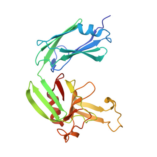
4JMX

Structure of LD transpeptidase LdtMt1 in complex with imipenem


Experimental Data Snapshot

  • Method: X-RAY DIFFRACTION
  • Resolution: 2.55 Å
  • R-Value Free: 0.233 
  • R-Value Work: 0.161 
  • R-Value Observed: 0.164 

wwPDB Validation   3D Report Full Report


Ligand Structure Quality Assessment 


This is version 1.1 of the entry. See complete history


Literature

Structures of free and inhibited forms of the L,D-transpeptidase LdtMt1 from Mycobacterium tuberculosis.

Correale, S.Ruggiero, A.Capparelli, R.Pedone, E.Berisio, R.

(2013) Acta Crystallogr D Biol Crystallogr 69: 1697-1706

  • DOI: https://doi.org/10.1107/S0907444913013085
  • Primary Citation of Related Structures:  
    4JMN, 4JMX

  • PubMed Abstract: 

    The modelling of peptidoglycan is responsible for key cellular processes in Mycobacterium tuberculosis such as cell growth, division and resuscitation from dormancy. The structure of M. tuberculosis peptidoglycan is atypical since it contains a majority of 3,3 cross-links synthesized by L,D-transpeptidases that replace the 4,3 cross-links formed by the D,D-transpeptidase activity of classical penicillin-binding proteins. Carbapenems inactivate these L,D-transpeptidases and in combination with clavulanic acid are bactericidal against extensively drug-resistant M. tuberculosis. Here, crystal structures of the L,D-transpeptidase LdtMt1 from M. tuberculosis in a ligand-free form and in complex with the carbapenem imipenem are reported. Elucidation of the structural features of LdtMt1 unveils analogies and differences between the two key transpeptidases of M. tuberculosis: LdtMt1 and LdtMt2. In addition, the structure of imipenem-inactivated LdtMt1 provides a detailed structural view of the interactions between a carbapenem drug and LdtMt1. By providing the key interactions in the binding of carbapenem to LdtMt1, this work will facilitate structure-guided discovery of L,D-transpeptidase inhibitors as novel antitubercular agents against drug-resistant M. tuberculosis.


  • Organizational Affiliation

    Institute of Biostructures and Bioimaging, CNR, Via Mezzocannone 16, 80134 Napoli, Italy.


Macromolecules
Find similar proteins by:  (by identity cutoff)  |  3D Structure
Entity ID: 1
MoleculeChains Sequence LengthOrganismDetailsImage
Probable L,D-transpeptidase LdtA220Mycobacterium tuberculosisMutation(s): 0 
Gene Names: ldtARv0116c
EC: 2.3.2
UniProt
Find proteins for O53638 (Mycobacterium tuberculosis (strain ATCC 25618 / H37Rv))
Explore O53638 
Go to UniProtKB:  O53638
Entity Groups  
Sequence Clusters30% Identity50% Identity70% Identity90% Identity95% Identity100% Identity
UniProt GroupO53638
Sequence Annotations
Expand
  • Reference Sequence
Small Molecules
Ligands 1 Unique
IDChains Name / Formula / InChI Key2D Diagram3D Interactions
IM2
Query on IM2

Download Ideal Coordinates CCD File 
B [auth A](5R)-5-[(1S,2R)-1-formyl-2-hydroxypropyl]-3-[(2-{[(E)-iminomethyl]amino}ethyl)sulfanyl]-4,5-dihydro-1H-pyrrole-2-carbox ylic acid
C12 H19 N3 O4 S
UACUABDJLSUFFC-IWSPIJDZSA-N
Binding Affinity Annotations 
IDSourceBinding Affinity
IM2 PDBBind:  4JMX Kd: 62.1 (nM) from 1 assay(s)
Experimental Data & Validation

Experimental Data

  • Method: X-RAY DIFFRACTION
  • Resolution: 2.55 Å
  • R-Value Free: 0.233 
  • R-Value Work: 0.161 
  • R-Value Observed: 0.164 
  • Space Group: P 43 21 2
Unit Cell:
Length ( Å )Angle ( ˚ )
a = 67.397α = 90
b = 67.397β = 90
c = 119.564γ = 90
Software Package:
Software NamePurpose
HKL-2000data collection
AMoREphasing
REFMACrefinement
HKL-2000data reduction
HKL-2000data scaling

Structure Validation

View Full Validation Report


Ligand Structure Quality Assessment 


Entry History 

Deposition Data

Revision History  (Full details and data files)

  • Version 1.0: 2013-10-23
    Type: Initial release
  • Version 1.1: 2024-10-16
    Changes: Data collection, Database references, Derived calculations, Structure summary