4JSZ | pdb_00004jsz

Benzenesulfonamide bound to hCAII H94C


Experimental Data Snapshot

  • Method: X-RAY DIFFRACTION
  • Resolution: 1.90 Å
  • R-Value Free: 
    0.230 (Depositor), 0.229 (DCC) 
  • R-Value Work: 
    0.166 (Depositor), 0.167 (DCC) 
  • R-Value Observed: 
    0.170 (Depositor) 

Starting Model: experimental
View more details

wwPDB Validation 3D Report Full Report

Validation slider image for 4JSZ

Ligand Structure Quality Assessment 


This is version 1.2 of the entry. See complete history

Literature

Metalloprotein-Inhibitor Binding: Human Carbonic Anhydrase II as a Model for Probing Metal-Ligand Interactions in a Metalloprotein Active Site.

Martin, D.P.Hann, Z.S.Cohen, S.M.

(2013) Inorg Chem 52: 12207-12215

  • DOI: https://doi.org/10.1021/ic400295f
  • Primary Citation Related Structures: 
    4JS6, 4JSA, 4JSS, 4JSW, 4JSZ

  • PubMed Abstract: 

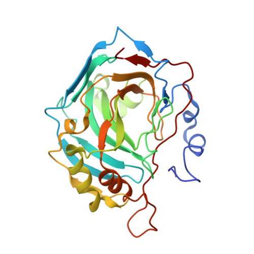
    An ever-increasing number of metalloproteins are being discovered that play essential roles in physiological processes. Inhibitors of these proteins have significant potential for the treatment of human disease, but clinical success of these compounds has been limited. Herein, zinc(II)-dependent metalloprotein inhibitors in clinical use are reviewed, and the potential for using novel metal-binding groups (MBGs) in the design of these inhibitors is discussed. By using human carbonic anhydrase II as a model system, the nuances of MBG-metal interactions in the context of a protein environment can be probed. Understanding how metal coordination influences inhibitor binding may help in the design of new therapeutics targeting metalloproteins.


  • Organizational Affiliation
    • Department of Chemistry and Biochemistry, University of California, San Diego , La Jolla, California 92093, United States.

Macromolecule Content 

  • Total Structure Weight: 29.63 kDa 
  • Atom Count: 2,216 
  • Modeled Residue Count: 257 
  • Deposited Residue Count: 260 
  • Unique protein chains: 1

Macromolecules

Find similar proteins by:|  3D Structure
Entity ID: 1
MoleculeChains  Sequence LengthOrganismDetailsImage
Carbonic anhydrase 2260Homo sapiensMutation(s): 1 
Gene Names: CA2
EC: 4.2.1.1 (PDB Primary Data), 4.2.1.69 (UniProt)
UniProt & NIH Common Fund Data Resources
Find proteins for P00918 (Homo sapiens)
Explore P00918 
Go to UniProtKB:  P00918
PHAROS:  P00918
GTEx:  ENSG00000104267 
Entity Groups
Sequence Clusters30% Identity50% Identity70% Identity90% Identity95% Identity100% Identity
UniProt GroupP00918
Sequence Annotations
Expand
Reference Sequence

Experimental Data & Validation

Experimental Data

  • Method: X-RAY DIFFRACTION
  • Resolution: 1.90 Å
  • R-Value Free:  0.230 (Depositor), 0.229 (DCC) 
  • R-Value Work:  0.166 (Depositor), 0.167 (DCC) 
  • R-Value Observed: 0.170 (Depositor) 
Space Group: P 1 21 1
Unit Cell:
Length ( Å )Angle ( ˚ )
a = 42.065α = 90
b = 41.345β = 104.27
c = 71.959γ = 90
Software Package:
Software NamePurpose
APEXdata collection
PHASERphasing
REFMACrefinement
APEXdata reduction

Structure Validation

View Full Validation Report


Ligand Structure Quality Assessment 


Entry History 

Deposition Data

Revision History  (Full details and data files)

  • Version 1.0: 2013-06-19
    Type: Initial release
  • Version 1.1: 2013-11-20
    Changes: Database references
  • Version 1.2: 2023-09-20
    Changes: Data collection, Database references, Derived calculations, Refinement description