Structural insight into the dimerization of human protein disulfide isomerase.
Bastos-Aristizabal, S., Kozlov, G., Gehring, K.(2014) Protein Sci 23: 618-626
- PubMed: 24549644 Search on PubMedSearch on PubMed Central
- DOI: https://doi.org/10.1002/pro.2444
- Primary Citation Related Structures:
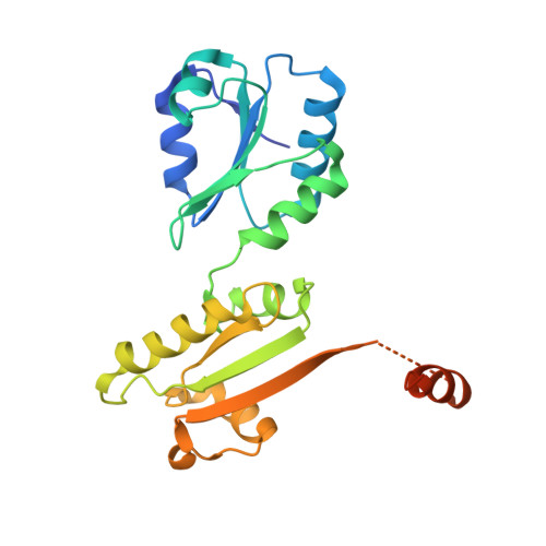
4JU5 - PubMed Abstract:
Protein disulfide isomerases (PDIs) are responsible for catalyzing the proper oxidation and isomerization of disulfide bonds of newly synthesized proteins in the endoplasmic reticulum (ER). Here, it is shown that human PDI (PDIA1) dimerizes in vivo and proposed that the dimerization of PDI has physiological relevance by autoregulating its activity. The crystal structure of the dimeric form of noncatalytic bb' domains of human PDIA1 determined to 2.3 Å resolution revealed that the formation of dimers occludes the substrate binding site and may function as a mechanism to regulate PDI activity in the ER.
- Department of Biochemistry, Groupe de recherche axé sur la structure des protéines, McGill University, 3649 Promenade Sir William Osler, Montréal, Québec, H3G 0B1, Canada.
Organizational Affiliation: