4KDT | pdb_00004kdt

Structure of an early native-like intermediate of beta2-microglobulin amyloidosis


Experimental Data Snapshot

  • Method: X-RAY DIFFRACTION
  • Resolution: 2.60 Å
  • R-Value Free: 
    0.274 (Depositor), 0.270 (DCC) 
  • R-Value Work: 
    0.226 (Depositor), 0.230 (DCC) 
  • R-Value Observed: 
    0.228 (Depositor) 

Starting Model: experimental
View more details

wwPDB Validation   3D Report Full Report


This is version 1.4 of the entry. See complete history


Literature

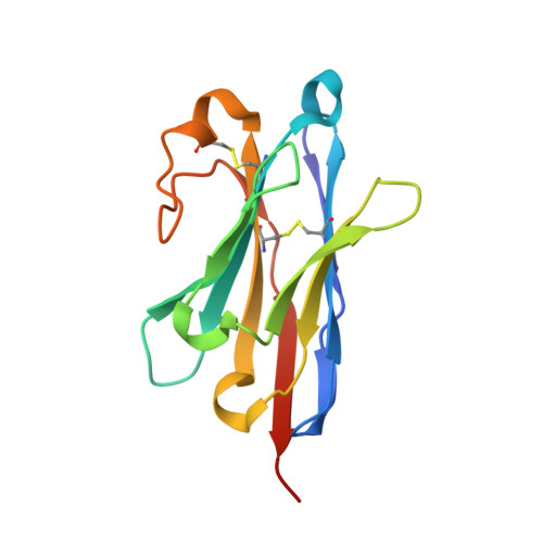
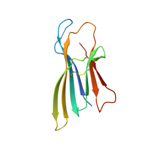
Structure of an early native-like intermediate of beta 2-microglobulin amyloidogenesis.

Vanderhaegen, S.Fislage, M.Domanska, K.Versees, W.Pardon, E.Bellotti, V.Steyaert, J.

(2013) Protein Sci 22: 1349-1357

  • DOI: https://doi.org/10.1002/pro.2321
  • Primary Citation of Related Structures:  
    4KDT

  • PubMed Abstract: 

    To investigate early intermediates of β2-microglobulin (β2m) amyloidogenesis, we solved the structure of β2m containing the amyloidogenic Pro32Gly mutation by X-ray crystallography. One nanobody (Nb24) that efficiently blocks fibril elongation was used as a chaperone to co-crystallize the Pro32Gly β2m monomer under physiological conditions. The complex of P32G β2m with Nb24 reveals a trans peptide bond at position 32 of this amyloidogenic variant, whereas Pro32 adopts the cis conformation in the wild-type monomer, indicating that the cis to trans isomerization at Pro32 plays a critical role in the early onset of β2m amyloid formation.


  • Organizational Affiliation
    • Structural Biology Research Centre, VIB, Pleinlaan 2, 1050, Brussel, Belgium; Structural Biology Brussels, Vrije Universiteit Brussel, Pleinlaan 2, 1050, Brussel, Belgium.

Macromolecules
Find similar proteins by:  (by identity cutoff)  |  3D Structure
Entity ID: 1
MoleculeChains Sequence LengthOrganismDetailsImage
Nanobody24
A, B
134CamelidaeMutation(s): 0 
Entity Groups  
Sequence Clusters30% Identity50% Identity70% Identity90% Identity95% Identity100% Identity
Sequence Annotations
Expand
  • Reference Sequence
Find similar proteins by:  (by identity cutoff)  |  3D Structure
Entity ID: 2
MoleculeChains Sequence LengthOrganismDetailsImage
Beta-2-microglobulin
C, D
100Homo sapiensMutation(s): 1 
Gene Names: B2MCDABP0092HDCMA22P
UniProt & NIH Common Fund Data Resources
Find proteins for P61769 (Homo sapiens)
Explore P61769 
Go to UniProtKB:  P61769
PHAROS:  P61769
GTEx:  ENSG00000166710 
Entity Groups  
Sequence Clusters30% Identity50% Identity70% Identity90% Identity95% Identity100% Identity
UniProt GroupP61769
Sequence Annotations
Expand
  • Reference Sequence
Small Molecules
Ligands 2 Unique
IDChains Name / Formula / InChI Key2D Diagram3D Interactions
SO4
Query on SO4

Download Ideal Coordinates CCD File 
E [auth A]
F [auth A]
G [auth A]
H [auth B]
I [auth C]
E [auth A],
F [auth A],
G [auth A],
H [auth B],
I [auth C],
K [auth D]
SULFATE ION
O4 S
QAOWNCQODCNURD-UHFFFAOYSA-L
GOL
Query on GOL

Download Ideal Coordinates CCD File 
J [auth C],
L [auth D]
GLYCEROL
C3 H8 O3
PEDCQBHIVMGVHV-UHFFFAOYSA-N
Experimental Data & Validation

Experimental Data

  • Method: X-RAY DIFFRACTION
  • Resolution: 2.60 Å
  • R-Value Free:  0.274 (Depositor), 0.270 (DCC) 
  • R-Value Work:  0.226 (Depositor), 0.230 (DCC) 
  • R-Value Observed: 0.228 (Depositor) 
Space Group: P 42 21 2
Unit Cell:
Length ( Å )Angle ( ˚ )
a = 96.79α = 90
b = 96.79β = 90
c = 167.79γ = 90
Software Package:
Software NamePurpose
PHASERphasing
REFMACrefinement
XDSdata reduction
XDSdata scaling

Structure Validation

View Full Validation Report


Entry History 

Deposition Data

Revision History  (Full details and data files)

  • Version 1.0: 2013-08-28
    Type: Initial release
  • Version 1.1: 2013-10-23
    Changes: Database references
  • Version 1.2: 2017-11-15
    Changes: Refinement description
  • Version 1.3: 2023-09-20
    Changes: Data collection, Database references, Derived calculations, Refinement description
  • Version 1.4: 2024-11-20
    Changes: Structure summary