


Structure of cIAP1-BIR3 and inhibitor
Li, X., Wang, J., Condon, S.M., Shi, Y.To be published.
Experimental Data Snapshot
Starting Model: experimental
View more details
Entity ID: 1 | |||||
|---|---|---|---|---|---|
| Molecule | Chains | Sequence Length | Organism | Details | Image |
| Baculoviral IAP repeat-containing protein 2 | 103 | Homo sapiens | Mutation(s): 0 Gene Names: BIRC2, API1, IAP2, MIHB, RNF48 EC: 6.3.2 (PDB Primary Data), 2.3.2.27 (UniProt) | 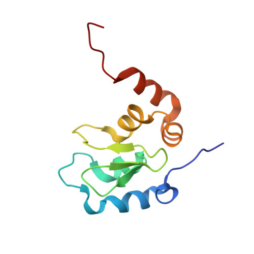 | |
UniProt & NIH Common Fund Data Resources | |||||
PHAROS: Q13490 GTEx: ENSG00000110330 | |||||
Entity Groups | |||||
| Sequence Clusters | 30% Identity50% Identity70% Identity90% Identity95% Identity100% Identity | ||||
| UniProt Group | Q13490 | ||||
Sequence AnnotationsExpand | |||||
Reference Sequence | |||||
| Ligands 3 Unique | |||||
|---|---|---|---|---|---|
| ID | Chains | Name / Formula / InChI Key | 2D Diagram | 3D Interactions | |
| 436 Download:Ideal Coordinates CCD File | C [auth A] | (2S)-N-{(2R)-1-[(2R,4S)-2-{[6,6'-difluoro-3'-({(2R,4S)-4-hydroxy-1-[(2S)-2-{[(2S)-2-(methylamino)propanoyl]amino}butanoyl]pyrrolidin-2-yl}methyl)-1H,1'H-2,2'-biindol-3-yl]methyl}-4-hydroxypyrrolidin-1-yl]-1-oxobutan-2-yl}-2-(methylamino)propanamide C42 H56 F2 N8 O6 PKWRMUKBEYJEIX-MNBKFCNDSA-N |  | ||
| PO4 Download:Ideal Coordinates CCD File | D [auth A] | PHOSPHATE ION O4 P NBIIXXVUZAFLBC-UHFFFAOYSA-K |  | ||
| ZN Download:Ideal Coordinates CCD File | B [auth A] | ZINC ION Zn PTFCDOFLOPIGGS-UHFFFAOYSA-N |  | ||
| Length ( Å ) | Angle ( ˚ ) |
|---|---|
| a = 67.714 | α = 90 |
| b = 67.714 | β = 90 |
| c = 66.913 | γ = 120 |
| Software Name | Purpose |
|---|---|
| PHENIX | refinement |


