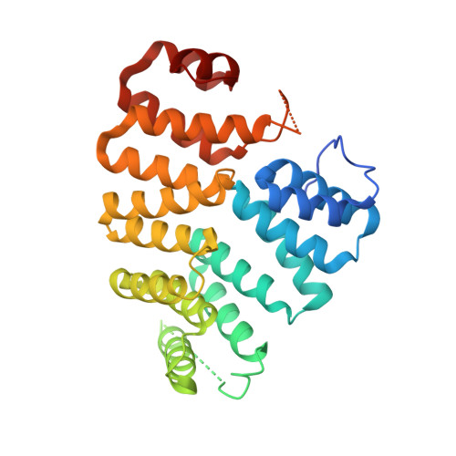
4KYO

Alanine-glyoxylate aminotransferase variant K390A in complex with the TPR domain of human Pex5p


Experimental Data Snapshot

  • Method: X-RAY DIFFRACTION
  • Resolution: 2.20 Å
  • R-Value Free: 0.218 
  • R-Value Work: 0.175 
  • R-Value Observed: 0.177 

Starting Model: experimental
View more details

wwPDB Validation   3D Report Full Report


This is version 1.2 of the entry. See complete history


Literature

Ligand-Induced Compaction of the PEX5 Receptor-Binding Cavity Impacts Protein Import Efficiency into Peroxisomes.

Fodor, K.Wolf, J.Reglinski, K.Passon, D.M.Lou, Y.Schliebs, W.Erdmann, R.Wilmanns, M.

(2015) Traffic 16: 85-98

  • DOI: https://doi.org/10.1111/tra.12238
  • Primary Citation of Related Structures:  
    4KXK, 4KYO

  • PubMed Abstract: 

    Peroxisomes entirely rely on the import of their proteome across the peroxisomal membrane. Recognition efficiencies of peroxisomal proteins vary by more than 1000-fold, but the molecular rationale behind their subsequent differential import and sorting has remained enigmatic. Using the protein cargo alanine-glyoxylate aminotransferase as a model, an unexpected increase from 34 to 80% in peroxisomal import efficiency of a single-residue mutant has been discovered. By high-resolution structural analysis, we found that it is the recognition receptor PEX5 that adapts its conformation for high-affinity binding rather than the cargo protein signal motif as previously thought. During receptor recognition, the binding cavity of the receptor shrinks to one third of its original volume. This process is impeded in the wild-type protein cargo because of a bulky side chain within the recognition motif, which blocks contraction of the PEX5 binding cavity. Our data provide a new insight into direct protein import efficiency by removal rather than by addition of an apparent specific sequence signature that is generally applicable to peroxisomal matrix proteins and to other receptor recognition processes.


  • Organizational Affiliation

    Hamburg Unit, European Molecular Biology Laboratory Hamburg Unit, Hamburg, Germany.


Macromolecules
Find similar proteins by:  (by identity cutoff)  |  3D Structure
Entity ID: 1
MoleculeChains Sequence LengthOrganismDetailsImage
Serine-pyruvate aminotransferaseA,
B [auth C]
394Homo sapiensMutation(s): 1 
Gene Names: AGXTAGT1SPAT
EC: 2.6.1.51 (PDB Primary Data), 2.6.1.44 (PDB Primary Data)
UniProt & NIH Common Fund Data Resources
Find proteins for P21549 (Homo sapiens)
Explore P21549 
Go to UniProtKB:  P21549
PHAROS:  P21549
GTEx:  ENSG00000172482 
Entity Groups  
Sequence Clusters30% Identity50% Identity70% Identity90% Identity95% Identity100% Identity
UniProt GroupP21549
Sequence Annotations
Expand
  • Reference Sequence
Find similar proteins by:  (by identity cutoff)  |  3D Structure
Entity ID: 2
MoleculeChains Sequence LengthOrganismDetailsImage
Peroxisomal targeting signal 1 receptorC [auth B],
D
328Homo sapiensMutation(s): 0 
Gene Names: PEX5PXR1
UniProt & NIH Common Fund Data Resources
Find proteins for P50542 (Homo sapiens)
Explore P50542 
Go to UniProtKB:  P50542
PHAROS:  P50542
GTEx:  ENSG00000139197 
Entity Groups  
Sequence Clusters30% Identity50% Identity70% Identity90% Identity95% Identity100% Identity
UniProt GroupP50542
Sequence Annotations
Expand
  • Reference Sequence
Small Molecules
Ligands 3 Unique
IDChains Name / Formula / InChI Key2D Diagram3D Interactions
BTB
Query on BTB

Download Ideal Coordinates CCD File 
G [auth A],
M [auth C]
2-[BIS-(2-HYDROXY-ETHYL)-AMINO]-2-HYDROXYMETHYL-PROPANE-1,3-DIOL
C8 H19 N O5
OWMVSZAMULFTJU-UHFFFAOYSA-N
SO4
Query on SO4

Download Ideal Coordinates CCD File 
E [auth A],
F [auth A]
SULFATE ION
O4 S
QAOWNCQODCNURD-UHFFFAOYSA-L
BME
Query on BME

Download Ideal Coordinates CCD File 
H [auth A]
I [auth A]
J [auth A]
K [auth A]
L [auth A]
H [auth A],
I [auth A],
J [auth A],
K [auth A],
L [auth A],
N [auth C],
O [auth C],
P [auth C],
Q [auth C],
R [auth C],
S [auth B],
T [auth D]
BETA-MERCAPTOETHANOL
C2 H6 O S
DGVVWUTYPXICAM-UHFFFAOYSA-N
Experimental Data & Validation

Experimental Data

  • Method: X-RAY DIFFRACTION
  • Resolution: 2.20 Å
  • R-Value Free: 0.218 
  • R-Value Work: 0.175 
  • R-Value Observed: 0.177 
  • Space Group: P 1
Unit Cell:
Length ( Å )Angle ( ˚ )
a = 57.556α = 87.23
b = 74.83β = 83.41
c = 91.751γ = 89.83
Software Package:
Software NamePurpose
DNAdata collection
PHASERphasing
REFMACrefinement
MOSFLMdata reduction
SCALAdata scaling

Structure Validation

View Full Validation Report


Entry History 

Deposition Data

Revision History  (Full details and data files)

  • Version 1.0: 2014-11-19
    Type: Initial release
  • Version 1.1: 2015-01-14
    Changes: Database references
  • Version 1.2: 2023-09-20
    Changes: Data collection, Database references, Derived calculations, Refinement description