4L2B | pdb_00004l2b

X-ray structure of the C57S mutant of the iron superoxide dismutase from Pseudoalteromonas haloplanktis


Experimental Data Snapshot

  • Method: X-RAY DIFFRACTION
  • Resolution: 1.97 Å
  • R-Value Free: 
    0.240 (Depositor), 0.249 (DCC) 
  • R-Value Work: 
    0.198 (Depositor), 0.205 (DCC) 
  • R-Value Observed: 
    0.198 (Depositor) 

wwPDB Validation 3D Report Full Report

Validation slider image for 4L2B

This is version 2.1 of the entry. See complete history

Literature

Structural and denaturation studies of two mutants of a cold adapted superoxide dismutase point to the importance of electrostatic interactions in protein stability.

Merlino, A.Russo Krauss, I.Castellano, I.Ruocco, M.R.Capasso, A.De Vendittis, E.Rossi, B.Sica, F.

(2014) Biochim Biophys Acta 1844: 632-640

  • DOI: https://doi.org/10.1016/j.bbapap.2014.01.007
  • Primary Citation Related Structures: 
    4L2A, 4L2B, 4L2C, 4L2D

  • PubMed Abstract: 

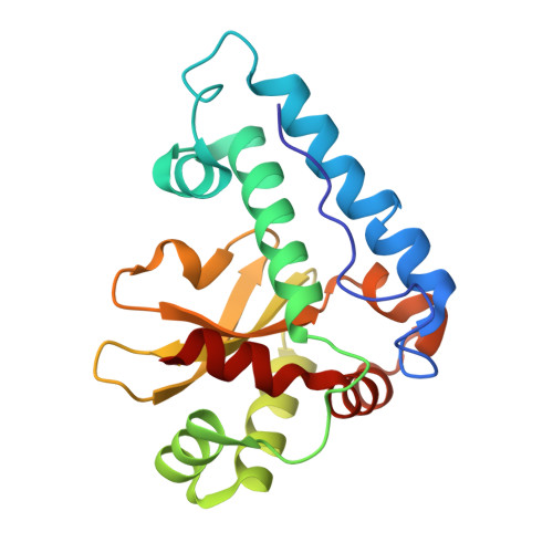
    A peculiar feature of the psychrophilic iron superoxide dismutase from Pseudoalteromonas haloplanktis (PhSOD) is the presence in its amino acid sequence of a reactive cysteine (Cys57). To define the role of this residue, a structural characterization of the effect of two PhSOD mutations, C57S and C57R, was performed. Thermal and denaturant-induced unfolding of wild type and mutant PhSOD followed by circular dichroism and fluorescence studies revealed that C→R substitution alters the thermal stability and the resistance against denaturants of the enzyme, whereas C57S only alters the stability of the protein against urea. The crystallographic data on the C57R mutation suggest an involvement of the Arg side chain in the formation of salt bridges on protein surface. These findings support the hypothesis that the thermal resistance of PhSOD relies on optimization of charge-charge interactions on its surface. Our study contributes to a deeper understanding of the denaturation mechanism of superoxide dismutases, suggesting the presence of a structural dimeric intermediate between the native state and the unfolded state. This hypothesis is supported by the crystalline and solution data on the reduced form of the enzyme.


  • Organizational Affiliation
    • Dipartimento di Scienze Chimiche, Università di Napoli "Federico II", Complesso Universitario di Monte Sant'Angelo, I-80126 Napoli, Italy; Istituto di Biostrutture e Bioimmagini, CNR, Via Mezzocannone 16, I-80134 Napoli, Italy.

Macromolecule Content 

  • Total Structure Weight: 43.3 kDa 
  • Atom Count: 3,473 
  • Modeled Residue Count: 384 
  • Deposited Residue Count: 384 
  • Unique protein chains: 1

Macromolecules

Find similar proteins by:|  3D Structure
Entity ID: 1
MoleculeChains  Sequence LengthOrganismDetailsImage
Superoxide dismutase [Fe]
A, B
192Pseudoalteromonas haloplanktisMutation(s): 1 
Gene Names: sodBPSHAa1215
EC: 1.15.1.1
UniProt
Find proteins for P84612 (Pseudoalteromonas translucida (strain TAC 125))
Explore P84612 
Go to UniProtKB:  P84612
Entity Groups
Sequence Clusters30% Identity50% Identity70% Identity90% Identity95% Identity100% Identity
UniProt GroupP84612
Sequence Annotations
Expand
Reference Sequence

Oligosaccharides

Help  
Entity ID: 2
MoleculeChains Length2D Diagram GlycosylationD Interactions
alpha-D-glucopyranose-(1-1)-alpha-D-glucopyranose
C, D
2N/A
Glycosylation Resources
GlyTouCan: G92130SN
GlyCosmos: G92130SN

Biologically Interesting Molecules (External Reference) 

1 Unique

Experimental Data & Validation

Experimental Data

  • Method: X-RAY DIFFRACTION
  • Resolution: 1.97 Å
  • R-Value Free:  0.240 (Depositor), 0.249 (DCC) 
  • R-Value Work:  0.198 (Depositor), 0.205 (DCC) 
  • R-Value Observed: 0.198 (Depositor) 
Space Group: P 1 21 1
Unit Cell:
Length ( Å )Angle ( ˚ )
a = 46.717α = 90
b = 103.436β = 107.97
c = 50.535γ = 90
Software Package:
Software NamePurpose
CrystalCleardata collection
PHASESphasing
CNSrefinement
HKL-2000data reduction
HKL-2000data scaling

Structure Validation

View Full Validation Report


Entry History 

Deposition Data

Revision History  (Full details and data files)

  • Version 1.0: 2014-02-26
    Type: Initial release
  • Version 2.0: 2020-07-29
    Type: Remediation
    Reason: Carbohydrate remediation
    Changes: Atomic model, Data collection, Database references, Derived calculations, Non-polymer description, Structure summary
  • Version 2.1: 2024-02-28
    Changes: Data collection, Database references, Structure summary