4MDQ

Structure of a novel submicromolar MDM2 inhibitor


Experimental Data Snapshot

  • Method: X-RAY DIFFRACTION
  • Resolution: 2.12 Å
  • R-Value Free: 0.221 
  • R-Value Work: 0.185 
  • R-Value Observed: 0.186 

wwPDB Validation   3D Report Full Report


Ligand Structure Quality Assessment 


This is version 1.3 of the entry. See complete history


Literature

Transient Protein States in Designing Inhibitors of the MDM2-p53 Interaction.

Bista, M.Wolf, S.Khoury, K.Kowalska, K.Huang, Y.Wrona, E.Arciniega, M.Popowicz, G.M.Holak, T.A.Domling, A.

(2013) Structure 21: 2143-2151

  • DOI: https://doi.org/10.1016/j.str.2013.09.006
  • Primary Citation of Related Structures:  
    4MDN, 4MDQ

  • PubMed Abstract: 

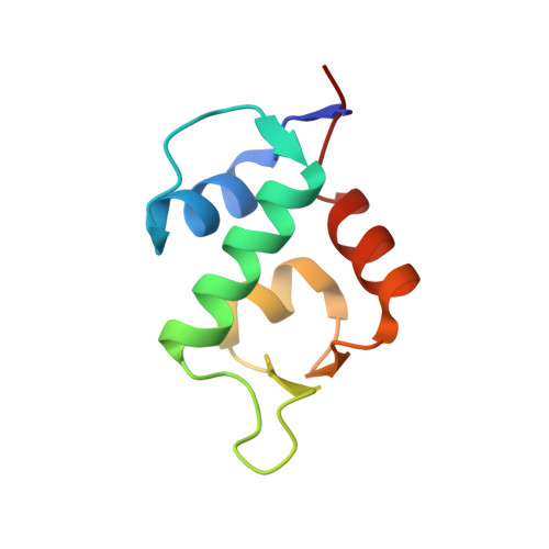
    Reactivation of p53 by release of the functional protein from its inhibition by MDM2 provides an efficient, nongenotoxic approach to a wide variety of cancers. We present the cocrystal structures of two complexes of MDM2 with inhibitors based on 6-chloroindole scaffolds. Both molecules bound to a distinct conformational state of MDM2 with nM-μM affinities. In contrast to other structurally characterized antagonists, which mimic three amino acids of p53 (Phe19, Trp23, and Leu26), the compounds induced an additional hydrophobic pocket on the MDM2 surface and unveiled a four-point binding mode. The enlarged interaction interface of the inhibitors resulted in extension of small molecules binding toward the "lid" segment of MDM2 (residues 19-23)--a nascent element that interferes with p53 binding. As supported by protein engineering and molecular dynamics studies, employing these unstable elements of MDM2 provides an efficient and yet unexplored alternative in development of MDM2-p53 association inhibitors.


  • Organizational Affiliation

    Max-Planck-Institute of Biochemistry, 82152 Martinsried, Germany.


Macromolecules
Find similar proteins by:  (by identity cutoff)  |  3D Structure
Entity ID: 1
MoleculeChains Sequence LengthOrganismDetailsImage
E3 ubiquitin-protein ligase Mdm286Homo sapiensMutation(s): 0 
Gene Names: MDM2
EC: 6.3.2
UniProt & NIH Common Fund Data Resources
Find proteins for Q00987 (Homo sapiens)
Explore Q00987 
Go to UniProtKB:  Q00987
PHAROS:  Q00987
GTEx:  ENSG00000135679 
Entity Groups  
Sequence Clusters30% Identity50% Identity70% Identity90% Identity95% Identity100% Identity
UniProt GroupQ00987
Sequence Annotations
Expand
  • Reference Sequence
Small Molecules
Ligands 1 Unique
IDChains Name / Formula / InChI Key2D Diagram3D Interactions
28W
Query on 28W

Download Ideal Coordinates CCD File 
B [auth A],
C [auth A]
3-[(1R)-2-(benzylamino)-1-{[(2S)-1-(hydroxyamino)-4-methyl-1-oxopentan-2-yl]amino}-2-oxoethyl]-6-chloro-N-hydroxy-1H-indole-2-carboxamide
C24 H28 Cl N5 O5
VRQFWSWBTGHUMK-AZUAARDMSA-N
Binding Affinity Annotations 
IDSourceBinding Affinity
28W BindingDB:  4MDQ Ki: min: 900, max: 1200 (nM) from 2 assay(s)
Binding MOAD:  4MDQ Ki: 1200 (nM) from 1 assay(s)
PDBBind:  4MDQ Ki: 1200 (nM) from 1 assay(s)
Experimental Data & Validation

Experimental Data

  • Method: X-RAY DIFFRACTION
  • Resolution: 2.12 Å
  • R-Value Free: 0.221 
  • R-Value Work: 0.185 
  • R-Value Observed: 0.186 
  • Space Group: P 65 2 2
Unit Cell:
Length ( Å )Angle ( ˚ )
a = 53.52α = 90
b = 53.52β = 90
c = 122.27γ = 120
Software Package:
Software NamePurpose
REFMACrefinement
PDB_EXTRACTdata extraction

Structure Validation

View Full Validation Report


Ligand Structure Quality Assessment 


Entry History 

Deposition Data

Revision History  (Full details and data files)

  • Version 1.0: 2013-11-13
    Type: Initial release
  • Version 1.1: 2013-12-04
    Changes: Database references
  • Version 1.2: 2013-12-25
    Changes: Database references
  • Version 1.3: 2024-02-28
    Changes: Data collection, Database references, Derived calculations