


Crystal structure of SUMO E3 Ligase PIAS3
Hu, J., Dong, a., Li, Y., Tempel, W., Bountra, C., Arrowsmith, C.H., Edwards, A.M., Tong, Y., Structural Genomics Consortium (SGC)To be published.
Experimental Data Snapshot
Starting Model: experimental
View more details
wwPDB Validation 3D Report Full Report

Entity ID: 1 | |||||
|---|---|---|---|---|---|
| Molecule | Chains | Sequence Length | Organism | Details | Image |
| E3 SUMO-protein ligase PIAS3 | 374 | Homo sapiens | Mutation(s): 0 Gene Names: PIAS3 EC: 6.3.2 (PDB Primary Data), 2.3.2 (UniProt) | 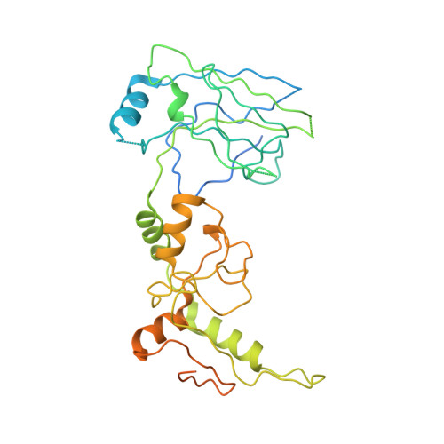 | |
UniProt & NIH Common Fund Data Resources | |||||
PHAROS: Q9Y6X2 GTEx: ENSG00000131788 | |||||
Entity Groups | |||||
| Sequence Clusters | 30% Identity50% Identity70% Identity90% Identity95% Identity100% Identity | ||||
| UniProt Group | Q9Y6X2 | ||||
Sequence AnnotationsExpand | |||||
Reference Sequence | |||||
| Ligands 3 Unique | |||||
|---|---|---|---|---|---|
| ID | Chains | Name / Formula / InChI Key | 2D Diagram | 3D Interactions | |
| ZN Download:Ideal Coordinates CCD File | E [auth A], JA [auth D], Q [auth B], Y [auth C] | ZINC ION Zn PTFCDOFLOPIGGS-UHFFFAOYSA-N |  | ||
| CL Download:Ideal Coordinates CCD File | F [auth A], Z [auth C] | CHLORIDE ION Cl VEXZGXHMUGYJMC-UHFFFAOYSA-M |  | ||
| UNX Download:Ideal Coordinates CCD File | AA [auth C] BA [auth C] CA [auth C] DA [auth C] EA [auth C] | UNKNOWN ATOM OR ION X |  | ||
| Length ( Å ) | Angle ( ˚ ) |
|---|---|
| a = 55.45 | α = 83.08 |
| b = 85.44 | β = 86.57 |
| c = 89.53 | γ = 86.14 |
| Software Name | Purpose |
|---|---|
| DENZO | data reduction |
| SCALEPACK | data scaling |
| REFMAC | refinement |
| PDB_EXTRACT | data extraction |
| JBluIce-EPICS | data collection |
| HKL-3000 | data reduction |
| MOLREP | phasing |
| BUSTER | refinement |
| Coot | model building |


