4NE9

PCSK9 in complex with LDLR peptide


Experimental Data Snapshot

  • Method: X-RAY DIFFRACTION
  • Resolution: 2.60 Å
  • R-Value Free: 0.211 
  • R-Value Work: 0.190 
  • R-Value Observed: 0.191 

wwPDB Validation   3D Report Full Report


Ligand Structure Quality Assessment 


This is version 2.0 of the entry. See complete history


Literature

Design and synthesis of truncated EGF-A peptides that restore LDL-R recycling in the presence of PCSK9 in vitro.

Schroeder, C.I.Swedberg, J.E.Withka, J.M.Rosengren, K.J.Akcan, M.Clayton, D.J.Daly, N.L.Cheneval, O.Borzilleri, K.A.Griffor, M.Stock, I.Colless, B.Walsh, P.Sunderland, P.Reyes, A.Dullea, R.Ammirati, M.Liu, S.McClure, K.F.Tu, M.Bhattacharya, S.K.Liras, S.Price, D.A.Craik, D.J.

(2014) Chem Biol 21: 284-294

  • DOI: https://doi.org/10.1016/j.chembiol.2013.11.014
  • Primary Citation of Related Structures:  
    2MG9, 4NE9

  • PubMed Abstract: 

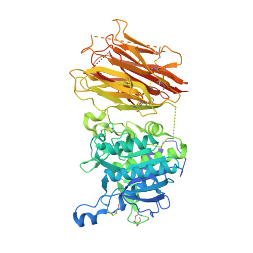
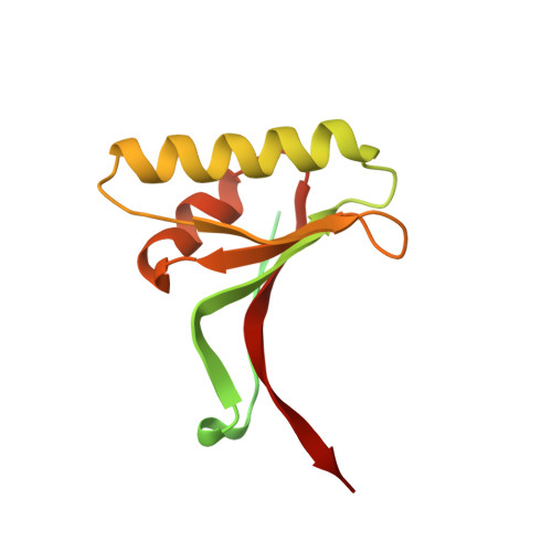
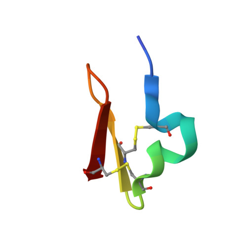
    Disrupting the binding interaction between proprotein convertase (PCSK9) and the epidermal growth factor-like domain A (EGF-A domain) in the low-density lipoprotein receptor (LDL-R) is a promising strategy to promote LDL-R recycling and thereby lower circulating cholesterol levels. In this study, truncated 26 amino acid EGF-A analogs were designed and synthesized, and their structures were analyzed in solution and in complex with PCSK9. The most potent peptide had an increased binding affinity for PCSK9 (KD = 0.6 μM) compared with wild-type EGF-A (KD = 1.2 μM), and the ability to increase LDL-R recycling in the presence of PCSK9 in a cell-based assay.


  • Organizational Affiliation

    Institute for Molecular Bioscience, The University of Queensland, Brisbane, 4072 QLD, Australia.


Macromolecules
Find similar proteins by:  (by identity cutoff)  |  3D Structure
Entity ID: 1
MoleculeChains Sequence LengthOrganismDetailsImage
Proprotein convertase subtilisin/kexin type 9
A, B
540Homo sapiensMutation(s): 2 
Gene Names: PCSK9NARC1PSEC0052
EC: 3.4.21
UniProt & NIH Common Fund Data Resources
Find proteins for Q8NBP7 (Homo sapiens)
Explore Q8NBP7 
Go to UniProtKB:  Q8NBP7
PHAROS:  Q8NBP7
GTEx:  ENSG00000169174 
Entity Groups  
Sequence Clusters30% Identity50% Identity70% Identity90% Identity95% Identity100% Identity
UniProt GroupQ8NBP7
Sequence Annotations
Expand
  • Reference Sequence
Find similar proteins by:  (by identity cutoff)  |  3D Structure
Entity ID: 2
MoleculeChains Sequence LengthOrganismDetailsImage
Proprotein convertase subtilisin/kexin type 9C,
E [auth P]
152Homo sapiensMutation(s): 0 
Gene Names: PCSK9NARC1PSEC0052
UniProt & NIH Common Fund Data Resources
Find proteins for Q8NBP7 (Homo sapiens)
Explore Q8NBP7 
Go to UniProtKB:  Q8NBP7
PHAROS:  Q8NBP7
GTEx:  ENSG00000169174 
Entity Groups  
Sequence Clusters30% Identity50% Identity70% Identity90% Identity95% Identity100% Identity
UniProt GroupQ8NBP7
Sequence Annotations
Expand
  • Reference Sequence
Find similar proteins by:  (by identity cutoff)  |  3D Structure
Entity ID: 3
MoleculeChains Sequence LengthOrganismDetailsImage
Low-density lipoprotein receptor26Homo sapiensMutation(s): 0 
Gene Names: LDLR
UniProt & NIH Common Fund Data Resources
Find proteins for P01130 (Homo sapiens)
Explore P01130 
Go to UniProtKB:  P01130
PHAROS:  P01130
GTEx:  ENSG00000130164 
Entity Groups  
Sequence Clusters30% Identity50% Identity70% Identity90% Identity95% Identity100% Identity
UniProt GroupP01130
Sequence Annotations
Expand
  • Reference Sequence
Oligosaccharides

Help

Entity ID: 4
MoleculeChains Length2D Diagram Glycosylation3D Interactions
alpha-L-fucopyranose-(1-6)-2-acetamido-2-deoxy-beta-D-glucopyranoseF [auth E]2N-Glycosylation
Glycosylation Resources
GlyTouCan:  G86851RC
GlyCosmos:  G86851RC
GlyGen:  G86851RC
Small Molecules
Ligands 2 Unique
IDChains Name / Formula / InChI Key2D Diagram3D Interactions
NAG
Query on NAG

Download Ideal Coordinates CCD File 
G [auth B]2-acetamido-2-deoxy-beta-D-glucopyranose
C8 H15 N O6
OVRNDRQMDRJTHS-FMDGEEDCSA-N
CA
Query on CA

Download Ideal Coordinates CCD File 
H [auth D]CALCIUM ION
Ca
BHPQYMZQTOCNFJ-UHFFFAOYSA-N
Experimental Data & Validation

Experimental Data

  • Method: X-RAY DIFFRACTION
  • Resolution: 2.60 Å
  • R-Value Free: 0.211 
  • R-Value Work: 0.190 
  • R-Value Observed: 0.191 
  • Space Group: P 21 21 21
Unit Cell:
Length ( Å )Angle ( ˚ )
a = 126.398α = 90
b = 131.392β = 90
c = 134.634γ = 90
Software Package:
Software NamePurpose
BUSTERrefinement

Structure Validation

View Full Validation Report


Ligand Structure Quality Assessment 


Entry History 

Deposition Data

  • Released Date: 2014-09-10 
  • Deposition Author(s): Liu, S.

Revision History  (Full details and data files)

  • Version 1.0: 2014-09-10
    Type: Initial release
  • Version 2.0: 2020-07-29
    Type: Remediation
    Reason: Carbohydrate remediation
    Changes: Atomic model, Data collection, Derived calculations, Structure summary