4NH1

Crystal structure of a heterotetrameric CK2 holoenzyme complex carrying the Andante-mutation in CK2beta and consistent with proposed models of autoinhibition and trans-autophosphorylation


Experimental Data Snapshot

  • Method: X-RAY DIFFRACTION
  • Resolution: 3.30 Å
  • R-Value Free: 0.251 
  • R-Value Work: 0.213 
  • R-Value Observed: 0.215 

wwPDB Validation   3D Report Full Report


Ligand Structure Quality Assessment 


This is version 1.2 of the entry. See complete history


Literature

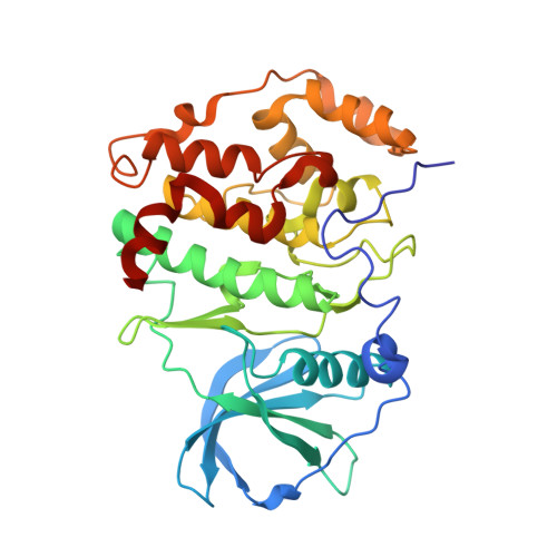
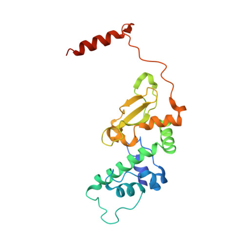
The Protein Kinase CK2(Andante) Holoenzyme Structure Supports Proposed Models of Autoregulation and Trans-Autophosphorylation

Schnitzler, A.Olsen, B.B.Issinger, O.-G.Niefind, K.

(2014) J Mol Biol 426: 1871-1882

  • DOI: https://doi.org/10.1016/j.jmb.2014.02.018
  • Primary Citation of Related Structures:  
    4NH1

  • PubMed Abstract: 

    Eukaryotic protein kinases are typically strictly controlled by second messenger binding, protein/protein interactions, dephosphorylations or similar processes. None of these regulatory mechanisms is known to work for protein kinase CK2 (former name "casein kinase 2"), an acidophilic and constitutively active eukaryotic protein kinase. CK2 predominantly exists as a heterotetrameric holoenzyme composed of two catalytic subunits (CK2α) complexed to a dimer of non-catalytic subunits (CK2β). One model of CK2 regulation was proposed several times independently by theoretical docking of the first CK2 holoenzyme structure. According to this model, the CK2 holoenzyme forms autoinhibitory aggregates correlated with trans-autophosphorylation and driven by the down-regulatory affinity between an acidic loop of CK2β and the positively charged substrate binding region of CK2α from a neighboring CK2 heterotetramer. Circular trimeric aggregates in which one-half of the CK2α chains show the predicted inhibitory proximity between those regions were detected within the crystal packing of the human CK2 holoenzyme. Here, we present further in vitro support of the "regulation-by-aggregation" model by an alternative crystal form in which CK2 tetramers are arranged as approximately linear aggregates coinciding essentially with the early predictions. In this assembly, the substrate binding region of every CK2α chain is blocked by a CK2β acidic loop from a neighboring tetramer. We found these crystals with CK2(Andante) that contains a CK2β variant mutated in a CK2α-contact helix and described to be responsible for a prolonged circadian rhythm in Drosophila. The increased propensity of CK2(Andante) to form aggregates with completely blocked active sites may contribute to this phenotype.


  • Organizational Affiliation

    Institut für Biochemie, Department für Chemie, Universität zu Köln, Otto-Fischer-Straße 12-14, D-50674 Köln, Germany.


Macromolecules
Find similar proteins by:  (by identity cutoff)  |  3D Structure
Entity ID: 1
MoleculeChains Sequence LengthOrganismDetailsImage
Casein kinase II subunit alpha
A, B
335Homo sapiensMutation(s): 0 
Gene Names: CSNK2A1
EC: 2.7.11.1
UniProt & NIH Common Fund Data Resources
Find proteins for P68400 (Homo sapiens)
Explore P68400 
Go to UniProtKB:  P68400
PHAROS:  P68400
GTEx:  ENSG00000101266 
Entity Groups  
Sequence Clusters30% Identity50% Identity70% Identity90% Identity95% Identity100% Identity
UniProt GroupP68400
Sequence Annotations
Expand
  • Reference Sequence
Find similar proteins by:  (by identity cutoff)  |  3D Structure
Entity ID: 2
MoleculeChains Sequence LengthOrganismDetailsImage
Casein kinase II subunit beta
C, D
215Homo sapiensMutation(s): 1 
Gene Names: CSNK2B
UniProt & NIH Common Fund Data Resources
Find proteins for P67870 (Homo sapiens)
Explore P67870 
Go to UniProtKB:  P67870
PHAROS:  P67870
GTEx:  ENSG00000204435 
Entity Groups  
Sequence Clusters30% Identity50% Identity70% Identity90% Identity95% Identity100% Identity
UniProt GroupP67870
Sequence Annotations
Expand
  • Reference Sequence
Small Molecules
Ligands 4 Unique
IDChains Name / Formula / InChI Key2D Diagram3D Interactions
ACP
Query on ACP

Download Ideal Coordinates CCD File 
G [auth A],
K [auth B]
PHOSPHOMETHYLPHOSPHONIC ACID ADENYLATE ESTER
C11 H18 N5 O12 P3
UFZTZBNSLXELAL-IOSLPCCCSA-N
GOL
Query on GOL

Download Ideal Coordinates CCD File 
J [auth B]GLYCEROL
C3 H8 O3
PEDCQBHIVMGVHV-UHFFFAOYSA-N
ZN
Query on ZN

Download Ideal Coordinates CCD File 
L [auth C],
M [auth D]
ZINC ION
Zn
PTFCDOFLOPIGGS-UHFFFAOYSA-N
MG
Query on MG

Download Ideal Coordinates CCD File 
E [auth A],
F [auth A],
H [auth B],
I [auth B]
MAGNESIUM ION
Mg
JLVVSXFLKOJNIY-UHFFFAOYSA-N
Experimental Data & Validation

Experimental Data

  • Method: X-RAY DIFFRACTION
  • Resolution: 3.30 Å
  • R-Value Free: 0.251 
  • R-Value Work: 0.213 
  • R-Value Observed: 0.215 
  • Space Group: C 1 2 1
Unit Cell:
Length ( Å )Angle ( ˚ )
a = 210.537α = 90
b = 57.441β = 118.54
c = 139.394γ = 90
Software Package:
Software NamePurpose
PHASERphasing
PHENIXrefinement
XDSdata reduction
XSCALEdata scaling

Structure Validation

View Full Validation Report


Ligand Structure Quality Assessment 


Entry History 

Deposition Data

Revision History  (Full details and data files)

  • Version 1.0: 2014-03-12
    Type: Initial release
  • Version 1.1: 2014-04-16
    Changes: Database references
  • Version 1.2: 2023-11-08
    Changes: Data collection, Database references, Derived calculations, Refinement description