Selective and Potent Morpholinone Inhibitors of the MDM2-p53 Protein-Protein Interaction.
Gonzalez, A.Z., Eksterowicz, J., Bartberger, M.D., Beck, H.P., Canon, J., Chen, A., Chow, D., Duquette, J., Fox, B.M., Fu, J., Huang, X., Houze, J.B., Jin, L., Li, Y., Li, Z., Ling, Y., Lo, M.C., Long, A.M., McGee, L.R., McIntosh, J., McMinn, D.L., Oliner, J.D., Osgood, T., Rew, Y., Saiki, A.Y., Shaffer, P., Wortman, S., Yakowec, P., Yan, X., Ye, Q., Yu, D., Zhao, X., Zhou, J., Olson, S.H., Medina, J.C., Sun, D.(2014) J Med Chem 57: 2472-2488
- PubMed: 24548297 Search on PubMed
- DOI: https://doi.org/10.1021/jm401767k
- Primary Citation Related Structures:
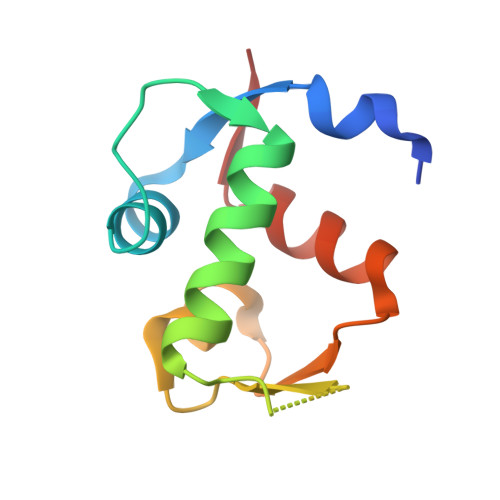
4OBA - PubMed Abstract:
We previously reported the discovery of AMG 232, a highly potent and selective piperidinone inhibitor of the MDM2-p53 interaction. Our continued search for potent and diverse analogues led to the discovery of novel morpholinone MDM2 inhibitors. This change to a morpholinone core has a significant impact on both potency and metabolic stability compared to the piperidinone series. Within this morpholinone series, AM-8735 emerged as an inhibitor with remarkable biochemical potency (HTRF IC50 = 0.4 nM) and cellular potency (SJSA-1 EdU IC50 = 25 nM), as well as pharmacokinetic properties. Compound 4 also shows excellent antitumor activity in the SJSA-1 osteosarcoma xenograft model with an ED50 of 41 mg/kg. Lead optimization toward the discovery of this inhibitor as well as key differences between the morpholinone and the piperidinone series will be described herein.
- Departments of †Therapeutic Discovery, ‡Pharmaceutics, and §Pharmacokinetics and Drug Metabolism, Amgen Inc. , 1120 Veterans Boulevard, South San Francisco, California 94080, United States.
Organizational Affiliation: