4OF9 | pdb_00004of9

Structure of K42N variant of sperm whale myoglobin


Experimental Data Snapshot

  • Method: X-RAY DIFFRACTION
  • Resolution: 1.24 Å
  • R-Value Free: 
    0.141 (Depositor), 0.139 (DCC) 
  • R-Value Work: 
    0.119 (Depositor), 0.118 (DCC) 
  • R-Value Observed: 
    0.120 (Depositor) 

Starting Model: experimental
View more details

wwPDB Validation 3D Report Full Report

Validation slider image for 4OF9

Ligand Structure Quality Assessment 


This is version 1.1 of the entry. See complete history

Literature

Structures of K42N and K42Y sperm whale myoglobins point to an inhibitory role of distal water in peroxidase activity.

Wang, C.Lovelace, L.L.Sun, S.Dawson, J.H.Lebioda, L.

(2014) Acta Crystallogr D Biol Crystallogr 70: 2833-2839

  • DOI: https://doi.org/10.1107/S1399004714017787
  • Primary Citation Related Structures: 
    4OF9, 4OOD

  • PubMed Abstract: 

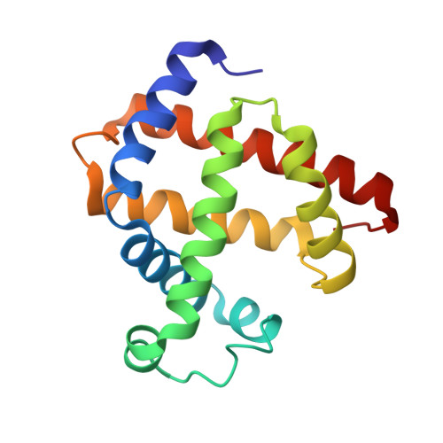
    Sperm whale myoglobin (Mb) functions as an oxygen-storage protein, but in the ferric state it possesses a weak peroxidase activity which enables it to carry out H2O2-dependent dehalogenation reactions. Hemoglobin/dehaloperoxidase from Amphitrite ornata (DHP) is a dual-function protein represented by two isoproteins DHP A and DHP B; its peroxidase activity is at least ten times stronger than that of Mb and plays a physiological role. The `DHP A-like' K42Y Mb mutant (K42Y) and the `DHP B-like' K42N mutant (K42N) were engineered in sperm whale Mb to mimic the extended heme environments of DHP A and DHP B, respectively. The peroxidase reaction rates increased ∼3.5-fold and ∼5.5-fold in K42Y and K42N versus Mb, respectively. The crystal structures of the K42Y and K42N mutants revealed that the substitutions at position 42 slightly elongate not only the distances between the distal His55 and the heme iron but also the hydrogen-bonding distances between His55 and the Fe-coordinated water. The enhanced peroxidase activity of K42Y and K42N thus might be attributed in part to the weaker binding of the axial water molecule that competes with hydrogen peroxide for the binding site at the heme in the ferric state. This is likely to be the mechanism by which the relationship `longer distal histidine to Fe distance - better peroxidase activity', which was previously proposed for heme proteins by Matsui et al. (1999) (J. Biol. Chem. 274, 2838-2844), works. Furthermore, positive cooperativity in K42N was observed when its dehaloperoxidase activity was measured as a function of the concentration of the substrate trichlorophenol. This serendipitously engineered cooperativity was rationalized by K42N dimerization through the formation of a dityrosine bond induced by excess H2O2.


  • Organizational Affiliation
    • Department of Chemistry and Biochemistry, University of South Carolina, Columbia, SC 29208, USA.

Macromolecule Content 

  • Total Structure Weight: 18.41 kDa 
  • Atom Count: 1,649 
  • Modeled Residue Count: 153 
  • Deposited Residue Count: 153 
  • Unique protein chains: 1

Macromolecules

Find similar proteins by:|  3D Structure
Entity ID: 1
MoleculeChains  Sequence LengthOrganismDetailsImage
Myoglobin153Physeter macrocephalusMutation(s): 1 
Gene Names: MB
EC: 1.11.1 (UniProt), 1.7 (UniProt)
UniProt
Find proteins for P02185 (Physeter macrocephalus)
Explore P02185 
Go to UniProtKB:  P02185
Entity Groups
Sequence Clusters30% Identity50% Identity70% Identity90% Identity95% Identity100% Identity
UniProt GroupP02185
Sequence Annotations
Expand
Reference Sequence

Small Molecules

Ligands 3 Unique
IDChains Name / Formula / InChI Key2D Diagram3D Interactions
HEM

Query on HEM


Download:Ideal Coordinates CCD File
B [auth A]PROTOPORPHYRIN IX CONTAINING FE
C34 H32 Fe N4 O4
KABFMIBPWCXCRK-RGGAHWMASA-L
SO4

Query on SO4


Download:Ideal Coordinates CCD File
C [auth A],
D [auth A],
E [auth A],
F [auth A]
SULFATE ION
O4 S
QAOWNCQODCNURD-UHFFFAOYSA-L
EDO

Query on EDO


Download:Ideal Coordinates CCD File
G [auth A],
H [auth A],
I [auth A]
1,2-ETHANEDIOL
C2 H6 O2
LYCAIKOWRPUZTN-UHFFFAOYSA-N

Experimental Data & Validation

Experimental Data

  • Method: X-RAY DIFFRACTION
  • Resolution: 1.24 Å
  • R-Value Free:  0.141 (Depositor), 0.139 (DCC) 
  • R-Value Work:  0.119 (Depositor), 0.118 (DCC) 
  • R-Value Observed: 0.120 (Depositor) 
Space Group: P 6
Unit Cell:
Length ( Å )Angle ( ˚ )
a = 90.304α = 90
b = 90.304β = 90
c = 45.314γ = 120
Software Package:
Software NamePurpose
AMoREphasing
REFMACrefinement
HKL-3000data reduction
SCALEPACKdata scaling

Structure Validation

View Full Validation Report


Ligand Structure Quality Assessment 


Entry History 

Deposition Data

Revision History  (Full details and data files)

  • Version 1.0: 2014-11-26
    Type: Initial release
  • Version 1.1: 2023-09-20
    Changes: Data collection, Database references, Derived calculations, Refinement description