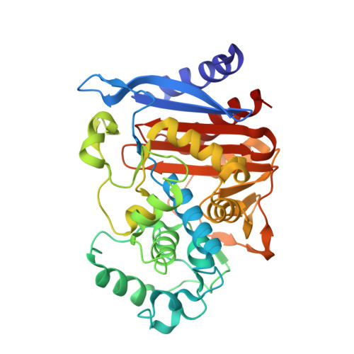
Substrate deconstruction and the nonadditivity of enzyme recognition.
Barelier, S., Cummings, J.A., Rauwerdink, A.M., Hitchcock, D.S., Farelli, J.D., Almo, S.C., Raushel, F.M., Allen, K.N., Shoichet, B.K.(2014) J Am Chem Soc 136: 7374-7382
- PubMed: 24791931
- DOI: https://doi.org/10.1021/ja501354q
- Primary Citation of Related Structures:
4OKP, 4OLD, 4OLG - PubMed Abstract:
Predicting substrates for enzymes of unknown function is a major postgenomic challenge. Substrate discovery, like inhibitor discovery, is constrained by our ability to explore chemotypes; it would be expanded by orders of magnitude if reactive sites could be probed with fragments rather than fully elaborated substrates, as is done for inhibitor discovery. To explore the feasibility of this approach, substrates of six enzymes from three different superfamilies were deconstructed into 41 overlapping fragments that were tested for activity or binding. Surprisingly, even those fragments containing the key reactive group had little activity, and most fragments did not bind measurably, until they captured most of the substrate features. Removing a single atom from a recognized substrate could often reduce catalytic recognition by 6 log-orders. To explore recognition at atomic resolution, the structures of three fragment complexes of the β-lactamase substrate cephalothin were determined by X-ray crystallography. Substrate discovery may be difficult to reduce to the fragment level, with implications for function discovery and for the tolerance of enzymes to metabolite promiscuity. Pragmatically, this study supports the development of libraries of fully elaborated metabolites as probes for enzyme function, which currently do not exist.
Organizational Affiliation:
Department of Pharmaceutical Chemistry, University of California - San Francisco , 1700 Fourth Street, Byers Hall, San Francisco, California 94158, United States.


