4OVN | pdb_00004ovn

Voltage-gated Sodium Channel 1.5 (Nav1.5) C-terminal domain in complex with Calmodulin poised for activation


Experimental Data Snapshot

  • Method: X-RAY DIFFRACTION
  • Resolution: 2.80 Å
  • R-Value Free: 
    0.282 (Depositor), 0.285 (DCC) 
  • R-Value Work: 
    0.211 (Depositor), 0.218 (DCC) 
  • R-Value Observed: 
    0.215 (Depositor) 

wwPDB Validation 3D Report Full Report

Validation slider image for 4OVN

This is version 1.6 of the entry. See complete history

Literature

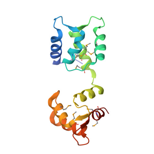
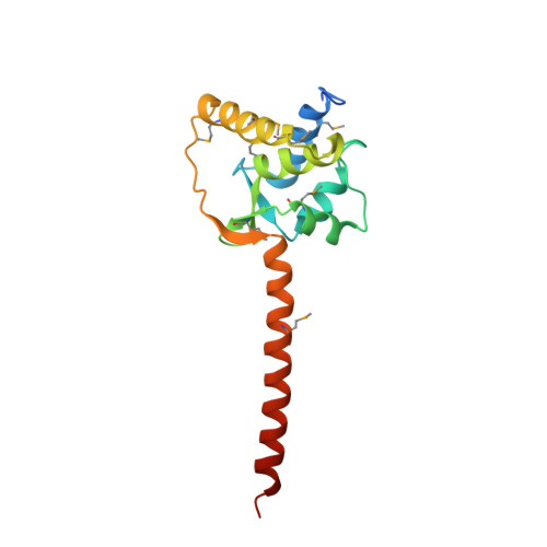
Regulation of the NaV1.5 cytoplasmic domain by calmodulin.

Gabelli, S.B.Boto, A.Kuhns, V.H.Bianchet, M.A.Farinelli, F.Aripirala, S.Yoder, J.Jakoncic, J.Tomaselli, G.F.Amzel, L.M.

(2014) Nat Commun 5: 5126

  • DOI: https://doi.org/10.1038/ncomms6126
  • Primary Citation Related Structures: 
    4OVN

  • PubMed Abstract: 

    Voltage-gated sodium channels (Na(v)) underlie the rapid upstroke of action potentials in excitable tissues. Binding of channel-interactive proteins is essential for controlling fast and long-term inactivation. In the structure of the complex of the carboxy-terminal portion of Na(v)1.5 (CTNa(v)1.5) with calmodulin (CaM)-Mg(2+) reported here, both CaM lobes interact with the CTNa(v)1.5. On the basis of the differences between this structure and that of an inactivated complex, we propose that the structure reported here represents a non-inactivated state of the CTNa(v), that is, the state that is poised for activation. Electrophysiological characterization of mutants further supports the importance of the interactions identified in the structure. Isothermal titration calorimetry experiments show that CaM binds to CTNa(v)1.5 with high affinity. The results of this study provide unique insights into the physiological activation and the pathophysiology of Na(v) channels.


  • Organizational Affiliation
    • 1] Structural Enzymology and Thermodynamics Group, Department of Biophysics and Biophysical Chemistry, Johns Hopkins University School of Medicine, 725 N Wolfe Street, WBSB 608, Baltimore, Maryland 21205, USA [2] Division of Cardiology, Department of Medicine, Johns Hopkins University School of Medicine, 720 Rutland Avenue, Ross Building 844, Baltimore, Maryland 21205, USA [3] Department of Oncology, Johns Hopkins University School of Medicine, Baltimore, Maryland 21287, USA.

Macromolecule Content 

  • Total Structure Weight: 180.93 kDa 
  • Atom Count: 11,784 
  • Modeled Residue Count: 1,458 
  • Deposited Residue Count: 1,530 
  • Unique protein chains: 2

Macromolecules

Find similar proteins by:|  3D Structure
Entity ID: 1
MoleculeChains  Sequence LengthOrganismDetailsImage
CalmodulinA,
C [auth B],
E [auth C],
G [auth D],
I [auth E]
149Homo sapiensMutation(s): 0 
Gene Names: 
CALM1CALMCAMCAM1CALM2CAM2CAMBCALM3CALML2CAM3...
CALM1CALMCAMCAM1CALM2CAM2CAMBCALM3CALML2CAM3CAMCCAMIII

UniProt & NIH Common Fund Data Resources
Find proteins for P0DP23 (Homo sapiens)
Explore P0DP23 
Go to UniProtKB:  P0DP23
PHAROS:  P0DP23
GTEx:  ENSG00000198668 
Entity Groups
Sequence Clusters30% Identity50% Identity70% Identity90% Identity95% Identity100% Identity
UniProt GroupP0DP23
Sequence Annotations
Expand
Reference Sequence
Find similar proteins by:|  3D Structure
Entity ID: 2
MoleculeChains  Sequence LengthOrganismDetailsImage
Sodium channel protein type 5 subunit alphaB [auth F],
D [auth G],
F [auth H],
H [auth I],
J
157Homo sapiensMutation(s): 0 
Gene Names: SCN5A
UniProt & NIH Common Fund Data Resources
Find proteins for Q14524 (Homo sapiens)
Explore Q14524 
Go to UniProtKB:  Q14524
PHAROS:  Q14524
GTEx:  ENSG00000183873 
Entity Groups
Sequence Clusters30% Identity50% Identity70% Identity90% Identity95% Identity100% Identity
UniProt GroupQ14524
Sequence Annotations
Expand
Reference Sequence

Small Molecules

Ligands 3 Unique
IDChains Name / Formula / InChI Key2D Diagram3D Interactions
SO4

Query on SO4


Download:Ideal Coordinates CCD File
BA [auth H],
IA [auth J],
O [auth A],
P [auth F],
U [auth G]
SULFATE ION
O4 S
QAOWNCQODCNURD-UHFFFAOYSA-L
PO4

Query on PO4


Download:Ideal Coordinates CCD File
T [auth B],
Z [auth C]
PHOSPHATE ION
O4 P
NBIIXXVUZAFLBC-UHFFFAOYSA-K
MG

Query on MG


Download:Ideal Coordinates CCD File
AA [auth C]
CA [auth D]
DA [auth D]
EA [auth D]
FA [auth D]
AA [auth C],
CA [auth D],
DA [auth D],
EA [auth D],
FA [auth D],
GA [auth E],
HA [auth E],
K [auth A],
L [auth A],
M [auth A],
N [auth A],
Q [auth B],
R [auth B],
S [auth B],
V [auth C],
W [auth C],
X [auth C],
Y [auth C]
MAGNESIUM ION
Mg
JLVVSXFLKOJNIY-UHFFFAOYSA-N
Modified Residues  1 Unique
IDChains TypeFormula2D DiagramParent
MSE
Query on MSE
A,
C [auth B],
E [auth C],
G [auth D],
I [auth E]
L-PEPTIDE LINKINGC5 H11 N O2 SeMET

Experimental Data & Validation

Experimental Data

  • Method: X-RAY DIFFRACTION
  • Resolution: 2.80 Å
  • R-Value Free:  0.282 (Depositor), 0.285 (DCC) 
  • R-Value Work:  0.211 (Depositor), 0.218 (DCC) 
  • R-Value Observed: 0.215 (Depositor) 
Space Group: P 1 21 1
Unit Cell:
Length ( Å )Angle ( ˚ )
a = 106.128α = 90
b = 99.006β = 106.11
c = 109.281γ = 90
Software Package:
Software NamePurpose
DENZOdata reduction
SCALEPACKdata scaling
REFMACrefinement

Structure Validation

View Full Validation Report


Entry History 

& Funding Information

Deposition Data


Funding OrganizationLocationGrant Number
National Institutes of Health/National Heart, Lung, and Blood Institute (NIH/NHLBI)United StatesRO1 HL050411-15A1

Revision History  (Full details and data files)

  • Version 1.0: 2014-12-03
    Type: Initial release
  • Version 1.1: 2014-12-10
    Changes: Derived calculations
  • Version 1.2: 2015-04-08
    Changes: Derived calculations
  • Version 1.3: 2017-09-27
    Changes: Author supporting evidence, Derived calculations, Refinement description, Source and taxonomy
  • Version 1.4: 2019-12-04
    Changes: Author supporting evidence
  • Version 1.5: 2023-12-27
    Changes: Data collection, Database references, Derived calculations
  • Version 1.6: 2024-10-30
    Changes: Structure summary