4P2H | pdb_00004p2h

Structure of human DNA polymerase complexed with N7MG in the template opposite to incoming non-hydrolyzable TTP with manganese in the active site


Experimental Data Snapshot

  • Method: X-RAY DIFFRACTION
  • Resolution: 1.99 Å
  • R-Value Free: 
    0.249 (Depositor), 0.249 (DCC) 
  • R-Value Work: 
    0.204 (Depositor), 0.204 (DCC) 
  • R-Value Observed: 
    0.206 (Depositor) 

Starting Model: experimental
View more details

wwPDB Validation 3D Report Full Report

Validation slider image for 4P2H

Ligand Structure Quality Assessment 


This is version 1.7 of the entry. See complete history

Literature

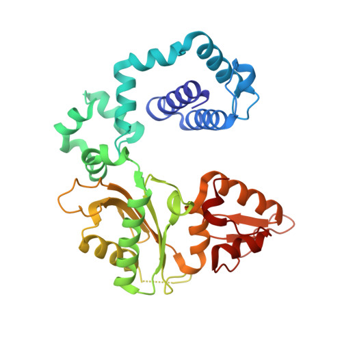
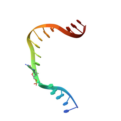
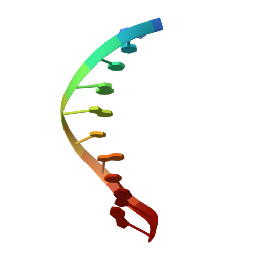
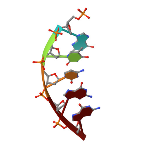
Transition-state destabilization reveals how human DNA polymerase beta proceeds across the chemically unstable lesion N7-methylguanine.

Koag, M.C.Kou, Y.Ouzon-Shubeita, H.Lee, S.

(2014) Nucleic Acids Res 42: 8755-8766

  • DOI: https://doi.org/10.1093/nar/gku554
  • Primary Citation Related Structures: 
    4O5C, 4O5E, 4O5K, 4P2H

  • PubMed Abstract: 

    N7-Methyl-2'-deoxyguanosine (m7dG) is the predominant lesion formed by methylating agents. A systematic investigation on the effect of m7dG on DNA replication has been difficult due to the chemical instability of m7dG. To gain insights into the m7dG effect, we employed a 2'-fluorine-mediated transition-state destabilzation strategy. Specifically, we determined kinetic parameters for dCTP insertion opposite a chemically stable m7dG analogue, 2'-fluoro-m7dG (Fm7dG), by human DNA polymerase β (polβ) and solved three X-ray structures of polβ in complex with the templating Fm7dG paired with incoming dCTP or dTTP analogues. The kinetic studies reveal that the templating Fm7dG slows polβ catalysis ∼ 300-fold, suggesting that m7dG in genomic DNA may impede replication by some DNA polymerases. The structural analysis reveals that Fm7dG forms a canonical Watson-Crick base pair with dCTP, but metal ion coordination is suboptimal for catalysis in the polβ-Fm7dG:dCTP complex, which partially explains the slow insertion of dCTP opposite Fm7dG by polβ. In addition, the polβ-Fm7dG:dTTP structure shows open protein conformations and staggered base pair conformations, indicating that N7-methylation of dG does not promote a promutagenic replication. Overall, the first systematic studies on the effect of m7dG on DNA replication reveal that polβ catalysis across m7dG is slow, yet highly accurate.


  • Organizational Affiliation
    • Division of Medicinal Chemistry, College of Pharmacy, The University of Texas at Austin, Austin, TX 78712, USA SeongminLee@austin.utexas.edu.

Macromolecule Content 

  • Total Structure Weight: 47.32 kDa 
  • Atom Count: 3,411 
  • Modeled Residue Count: 355 
  • Deposited Residue Count: 357 
  • Unique protein chains: 1
  • Unique nucleic acid chains: 3

Macromolecules


Find similar proteins by:|  3D Structure
Entity ID: 1
MoleculeChains  Sequence LengthOrganismDetailsImage
DNA polymerase beta326Homo sapiensMutation(s): 0 
Gene Names: POLB
EC: 2.7.7.7 (PDB Primary Data), 4.2.99.18 (UniProt), 4.2.99 (UniProt)
UniProt & NIH Common Fund Data Resources
Find proteins for P06746 (Homo sapiens)
Explore P06746 
Go to UniProtKB:  P06746
PHAROS:  P06746
GTEx:  ENSG00000070501 
Entity Groups
Sequence Clusters30% Identity50% Identity70% Identity90% Identity95% Identity100% Identity
UniProt GroupP06746
Sequence Annotations
Expand
Reference Sequence
Find similar nucleic acids by:  Sequence
Entity ID: 2
MoleculeChains LengthOrganismImage
DNA (5'-D(*CP*CP*GP*AP*CP*(FMG)P*TP*CP*GP*CP*AP*TP*CP*AP*GP*C)-3')B [auth T]16Homo sapiens
Sequence Annotations
Expand
Reference Sequence
Find similar nucleic acids by:  Sequence
Entity ID: 3
MoleculeChains LengthOrganismImage
DNA (5'-D(*GP*CP*TP*GP*AP*TP*GP*CP*GP*A)-3')C [auth P]10Homo sapiens
Sequence Annotations
Expand
Reference Sequence
Find similar nucleic acids by:  Sequence
Entity ID: 4
MoleculeChains LengthOrganismImage
DNA (5'-D(P*GP*TP*CP*GP*G)-3')5Homo sapiens
Sequence Annotations
Expand
Reference Sequence

Experimental Data & Validation

Experimental Data

  • Method: X-RAY DIFFRACTION
  • Resolution: 1.99 Å
  • R-Value Free:  0.249 (Depositor), 0.249 (DCC) 
  • R-Value Work:  0.204 (Depositor), 0.204 (DCC) 
  • R-Value Observed: 0.206 (Depositor) 
Space Group: P 1 21 1
Unit Cell:
Length ( Å )Angle ( ˚ )
a = 54.767α = 90
b = 79.167β = 106.18
c = 54.982γ = 90
Software Package:
Software NamePurpose
SCALEPACKdata scaling
PDB_EXTRACTdata extraction
PHENIXrefinement

Structure Validation

View Full Validation Report


Ligand Structure Quality Assessment 


Entry History 

& Funding Information

Deposition Data


Funding OrganizationLocationGrant Number
National Institutes of Health/National Institute of Environmental Health Sciences (NIH/NIEHS)United StatesES23101

Revision History  (Full details and data files)

  • Version 1.0: 2014-06-25
    Type: Initial release
  • Version 1.1: 2014-07-09
    Changes: Database references
  • Version 1.2: 2014-10-01
    Changes: Database references
  • Version 1.3: 2015-01-07
    Changes: Database references
  • Version 1.4: 2017-09-20
    Changes: Author supporting evidence, Database references, Derived calculations, Other, Refinement description, Source and taxonomy
  • Version 1.5: 2017-09-27
    Changes: Refinement description
  • Version 1.6: 2019-12-18
    Changes: Author supporting evidence
  • Version 1.7: 2023-09-27
    Changes: Data collection, Database references, Derived calculations, Refinement description