4P3A | pdb_00004p3a

Crystal structure of the mouse C5a anaphylatoxin


Experimental Data Snapshot

  • Method: X-RAY DIFFRACTION
  • Resolution: 1.40 Å
  • R-Value Free: 
    0.174 (Depositor), 0.181 (DCC) 
  • R-Value Work: 
    0.144 (Depositor), 0.155 (DCC) 
  • R-Value Observed: 
    0.146 (Depositor) 

Starting Model: experimental
View more details

wwPDB Validation 3D Report Full Report

Validation slider image for 4P3A

This is version 1.4 of the entry. See complete history

Literature

Structural and functional characterization of human and murine C5a anaphylatoxins.

Schatz-Jakobsen, J.A.Yatime, L.Larsen, C.Petersen, S.V.Klos, A.Andersen, G.R.

(2014) Acta Crystallogr D Biol Crystallogr 70: 1704-1717

  • DOI: https://doi.org/10.1107/S139900471400844X
  • Primary Citation Related Structures: 
    4P39, 4P3A, 4P3B

  • PubMed Abstract: 

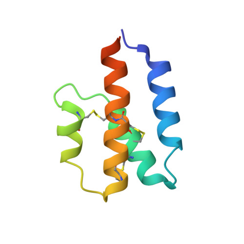
    Complement is an ancient part of the innate immune system that plays a pivotal role in protection against invading pathogens and helps to clear apoptotic and necrotic cells. Upon complement activation, a cascade of proteolytic events generates the complement effectors, including the anaphylatoxins C3a and C5a. Signalling through their cognate G-protein coupled receptors, C3aR and C5aR, leads to a wide range of biological events promoting inflammation at the site of complement activation. The function of anaphylatoxins is regulated by circulating carboxypeptidases that remove their C-terminal arginine residue, yielding C3a-desArg and C5a-desArg. Whereas human C3a and C3a-desArg adopt a canonical four-helix bundle fold, the conformation of human C5a-desArg has recently been described as a three-helix bundle. Here, the crystal structures of an antagonist version of human C5a, A8(Δ71-73), and of murine C5a and C5a-desArg are reported. Whereas A8(Δ71-73) adopts a three-helix bundle conformation similar to human C5a-desArg, the two murine proteins form a four-helix bundle. A cell-based functional assay reveals that murine C5a-desArg, in contrast to its human counterpart, exerts the same level of activition as murine C5a on its cognate receptor. The role of the different C5a conformations is discussed in relation to the differential activation of C5a receptors across species.


  • Organizational Affiliation
    • Department of Molecular Biology and Genetics, Aarhus University, Gustav Wieds Vej 10C, DK-8000 Aarhus, Denmark.

Macromolecule Content 

  • Total Structure Weight: 36.93 kDa 
  • Atom Count: 2,730 
  • Modeled Residue Count: 276 
  • Deposited Residue Count: 316 
  • Unique protein chains: 1

Macromolecules

Find similar proteins by:|  3D Structure
Entity ID: 1
MoleculeChains  Sequence LengthOrganismDetailsImage
Complement C5
A, B, C, D
79Mus musculusMutation(s): 0 
Gene Names: C5Hc
UniProt & NIH Common Fund Data Resources
Find proteins for P06684 (Mus musculus)
Explore P06684 
Go to UniProtKB:  P06684
IMPC:  MGI:96031
Entity Groups
Sequence Clusters30% Identity50% Identity70% Identity90% Identity95% Identity100% Identity
UniProt GroupP06684
Sequence Annotations
Expand
Reference Sequence

Small Molecules

Ligands 1 Unique
IDChains Name / Formula / InChI Key2D Diagram3D Interactions
FMT

Query on FMT


Download:Ideal Coordinates CCD File
E [auth A]
F [auth A]
G [auth A]
H [auth B]
I [auth B]
E [auth A],
F [auth A],
G [auth A],
H [auth B],
I [auth B],
J [auth B],
K [auth B],
L [auth B],
M [auth B],
N [auth B],
O [auth C],
P [auth C],
Q [auth D],
R [auth D],
S [auth D],
T [auth D]
FORMIC ACID
C H2 O2
BDAGIHXWWSANSR-UHFFFAOYSA-N

Experimental Data & Validation

Experimental Data

  • Method: X-RAY DIFFRACTION
  • Resolution: 1.40 Å
  • R-Value Free:  0.174 (Depositor), 0.181 (DCC) 
  • R-Value Work:  0.144 (Depositor), 0.155 (DCC) 
  • R-Value Observed: 0.146 (Depositor) 
Space Group: P 43
Unit Cell:
Length ( Å )Angle ( ˚ )
a = 54.95α = 90
b = 54.95β = 90
c = 117.43γ = 90
Software Package:
Software NamePurpose
XDSdata scaling
XSCALEdata scaling
PDB_EXTRACTdata extraction
PHASERphasing
PHENIXrefinement

Structure Validation

View Full Validation Report


Entry History 

Deposition Data

Revision History  (Full details and data files)

  • Version 1.0: 2014-06-11
    Type: Initial release
  • Version 1.1: 2014-06-25
    Changes: Database references
  • Version 1.2: 2017-09-27
    Changes: Database references, Derived calculations, Other, Refinement description, Source and taxonomy
  • Version 1.3: 2023-09-27
    Changes: Data collection, Database references, Refinement description
  • Version 1.4: 2024-10-23
    Changes: Structure summary