4P6X

Crystal Structure of cortisol-bound glucocorticoid receptor ligand binding domain


Experimental Data Snapshot

  • Method: X-RAY DIFFRACTION
  • Resolution: 2.50 Å
  • R-Value Free: 0.276 
  • R-Value Work: 0.246 
  • R-Value Observed: 0.248 

wwPDB Validation   3D Report Full Report


Ligand Structure Quality Assessment 


This is version 1.5 of the entry. See complete history


Literature

Structures and mechanism for the design of highly potent glucocorticoids.

He, Y.Yi, W.Suino-Powell, K.Zhou, X.E.Tolbert, W.D.Tang, X.Yang, J.Yang, H.Shi, J.Hou, L.Jiang, H.Melcher, K.Xu, H.E.

(2014) Cell Res 24: 713-726

  • DOI: https://doi.org/10.1038/cr.2014.52
  • Primary Citation of Related Structures:  
    4P6W, 4P6X

  • PubMed Abstract: 

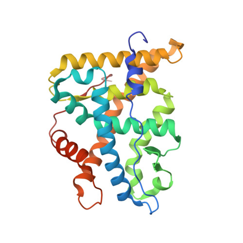
    The evolution of glucocorticoid drugs was driven by the demand of lowering the unwanted side effects, while keeping the beneficial anti-inflammatory effects. Potency is an important aspect of this evolution as many undesirable side effects are associated with use of high-dose glucocorticoids. The side effects can be minimized by highly potent glucocorticoids that achieve the same treatment effects at lower doses. This demand propelled the continuous development of synthetic glucocorticoids with increased potencies, but the structural basis of their potencies is poorly understood. To determine the mechanisms underlying potency, we solved the X-ray structures of the glucocorticoid receptor (GR) ligand-binding domain (LBD) bound to its endogenous ligand, cortisol, which has relatively low potency, and a highly potent synthetic glucocorticoid, mometasone furoate (MF). The cortisol-bound GR LBD revealed that the flexibility of the C1-C2 single bond in the steroid A ring is primarily responsible for the low affinity of cortisol to GR. In contrast, we demonstrate that the very high potency of MF is achieved by its C-17α furoate group completely filling the ligand-binding pocket, thus providing additional anchor contacts for high-affinity binding. A single amino acid in the ligand-binding pocket, Q642, plays a discriminating role in ligand potency between MF and cortisol. Structure-based design led to synthesis of several novel glucocorticoids with much improved potency and efficacy. Together, these results reveal key structural mechanisms of glucocorticoid potency and provide a rational basis for developing novel highly potent glucocorticoids.


  • Organizational Affiliation

    Laboratory of Structural Sciences, Van Andel Research Institute, Grand Rapids, MI 49503, USA.


Macromolecules
Find similar proteins by:  (by identity cutoff)  |  3D Structure
Entity ID: 1
MoleculeChains Sequence LengthOrganismDetailsImage
Glucocorticoid receptor
A, C, E, G, I
A, C, E, G, I, K
255Homo sapiensMutation(s): 7 
Gene Names: NR3C1GRL
UniProt & NIH Common Fund Data Resources
Find proteins for P04150 (Homo sapiens)
Explore P04150 
Go to UniProtKB:  P04150
PHAROS:  P04150
GTEx:  ENSG00000113580 
Entity Groups  
Sequence Clusters30% Identity50% Identity70% Identity90% Identity95% Identity100% Identity
UniProt GroupP04150
Sequence Annotations
Expand
  • Reference Sequence

Find similar proteins by:  Sequence   |   3D Structure  

Entity ID: 2
MoleculeChains Sequence LengthOrganismDetailsImage
Nuclear receptor coactivator 2
B, D, F, H, J
B, D, F, H, J, L
14Homo sapiensMutation(s): 0 
UniProt & NIH Common Fund Data Resources
Find proteins for Q15596 (Homo sapiens)
Explore Q15596 
Go to UniProtKB:  Q15596
PHAROS:  Q15596
GTEx:  ENSG00000140396 
Entity Groups  
Sequence Clusters30% Identity50% Identity70% Identity90% Identity95% Identity100% Identity
UniProt GroupQ15596
Sequence Annotations
Expand
  • Reference Sequence
Small Molecules
Binding Affinity Annotations 
IDSourceBinding Affinity
HCY Binding MOAD:  4P6X Ki: 91 (nM) from 1 assay(s)
BindingDB:  4P6X Ki: 30 (nM) from 1 assay(s)
Kd: 43 (nM) from 1 assay(s)
Experimental Data & Validation

Experimental Data

  • Method: X-RAY DIFFRACTION
  • Resolution: 2.50 Å
  • R-Value Free: 0.276 
  • R-Value Work: 0.246 
  • R-Value Observed: 0.248 
  • Space Group: P 61
Unit Cell:
Length ( Å )Angle ( ˚ )
a = 220.824α = 90
b = 220.824β = 90
c = 74.198γ = 120
Software Package:
Software NamePurpose
HKL-2000data scaling
PDB_EXTRACTdata extraction
PHASERphasing
PHENIXrefinement

Structure Validation

View Full Validation Report


Ligand Structure Quality Assessment 


Entry History & Funding Information

Deposition Data


Funding OrganizationLocationGrant Number
National Institutes of Health/National Institute of Diabetes and Digestive and Kidney Disease (NIH/NIDDK)United StatesDK066202
National Institutes of Health/National Institute of Diabetes and Digestive and Kidney Disease (NIH/NIDDK)United StatesDK071662
AAFUnited States2010 senior award

Revision History  (Full details and data files)

  • Version 1.0: 2014-04-16
    Type: Initial release
  • Version 1.1: 2014-05-07
    Changes: Database references
  • Version 1.2: 2014-07-16
    Changes: Database references
  • Version 1.3: 2017-09-27
    Changes: Author supporting evidence, Derived calculations, Other, Refinement description, Source and taxonomy, Structure summary
  • Version 1.4: 2019-12-25
    Changes: Author supporting evidence
  • Version 1.5: 2023-12-27
    Changes: Data collection, Database references, Refinement description