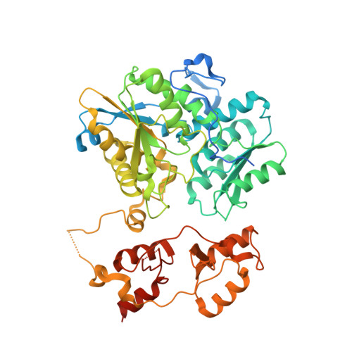
4PCU

Crystal structure of delta516-525 E201S human cystathionine beta-synthase with AdoMet


Experimental Data Snapshot

  • Method: X-RAY DIFFRACTION
  • Resolution: 3.58 Å
  • R-Value Free: 0.277 
  • R-Value Work: 0.257 
  • R-Value Observed: 0.258 

wwPDB Validation   3D Report Full Report


Ligand Structure Quality Assessment 


This is version 1.3 of the entry. See complete history


Literature

Structural insight into the molecular mechanism of allosteric activation of human cystathionine beta-synthase by S-adenosylmethionine.

Ereno-Orbea, J.Majtan, T.Oyenarte, I.Kraus, J.P.Martinez-Cruz, L.A.

(2014) Proc Natl Acad Sci U S A 111: E3845-E3852

  • DOI: https://doi.org/10.1073/pnas.1414545111
  • Primary Citation of Related Structures:  
    4PCU

  • PubMed Abstract: 

    Cystathionine β-synthase (CBS) is a heme-dependent and pyridoxal-5'-phosphate-dependent protein that controls the flux of sulfur from methionine to cysteine, a precursor of glutathione, taurine, and H2S. Deficiency of CBS activity causes homocystinuria, the most frequent disorder of sulfur amino acid metabolism. In contrast to CBSs from lower organisms, human CBS (hCBS) is allosterically activated by S-adenosylmethionine (AdoMet), which binds to the regulatory domain and triggers a conformational change that allows the protein to progress from the basal toward the activated state. The structural basis of the underlying molecular mechanism has remained elusive so far. Here, we present the structure of hCBS with bound AdoMet, revealing the activated conformation of the human enzyme. Binding of AdoMet triggers a conformational change in the Bateman module of the regulatory domain that favors its association with a Bateman module of the complementary subunit to form an antiparallel CBS module. Such an arrangement is very similar to that found in the constitutively activated insect CBS. In the presence of AdoMet, the autoinhibition exerted by the regulatory region is eliminated, allowing for improved access of substrates to the catalytic pocket. Based on the availability of both the basal and the activated structures, we discuss the mechanism of hCBS activation by AdoMet and the properties of the AdoMet binding site, as well as the responsiveness of the enzyme to its allosteric regulator. The structure described herein paves the way for the rational design of compounds modulating hCBS activity and thus transsulfuration, redox status, and H2S biogenesis.


  • Organizational Affiliation

    Structural Biology Unit, Center for Cooperative Research in Biosciences (CIC bioGUNE), Technology Park of Bizkaia, 48160 Derio, Spain; and.


Macromolecules
Find similar proteins by:  (by identity cutoff)  |  3D Structure
Entity ID: 1
MoleculeChains Sequence LengthOrganismDetailsImage
Cystathionine beta-synthase
A, B
549Homo sapiensMutation(s): 1 
Gene Names: CBS
EC: 4.2.1.22
UniProt & NIH Common Fund Data Resources
Find proteins for P35520 (Homo sapiens)
Explore P35520 
Go to UniProtKB:  P35520
PHAROS:  P35520
GTEx:  ENSG00000160200 
Entity Groups  
Sequence Clusters30% Identity50% Identity70% Identity90% Identity95% Identity100% Identity
UniProt GroupP35520
Sequence Annotations
Expand
  • Reference Sequence
Small Molecules
Ligands 3 Unique
IDChains Name / Formula / InChI Key2D Diagram3D Interactions
HEM
Query on HEM

Download Ideal Coordinates CCD File 
C [auth A],
F [auth B]
PROTOPORPHYRIN IX CONTAINING FE
C34 H32 Fe N4 O4
KABFMIBPWCXCRK-RGGAHWMASA-L
SAM
Query on SAM

Download Ideal Coordinates CCD File 
E [auth A],
H [auth B]
S-ADENOSYLMETHIONINE
C15 H22 N6 O5 S
MEFKEPWMEQBLKI-FCKMPRQPSA-N
PLP
Query on PLP

Download Ideal Coordinates CCD File 
D [auth A],
G [auth B]
PYRIDOXAL-5'-PHOSPHATE
C8 H10 N O6 P
NGVDGCNFYWLIFO-UHFFFAOYSA-N
Experimental Data & Validation

Experimental Data

  • Method: X-RAY DIFFRACTION
  • Resolution: 3.58 Å
  • R-Value Free: 0.277 
  • R-Value Work: 0.257 
  • R-Value Observed: 0.258 
  • Space Group: P 31 2 1
Unit Cell:
Length ( Å )Angle ( ˚ )
a = 141.354α = 90
b = 141.354β = 90
c = 108.528γ = 120
Software Package:
Software NamePurpose
PHENIXrefinement
PDB_EXTRACTdata extraction
XSCALEdata scaling

Structure Validation

View Full Validation Report


Ligand Structure Quality Assessment 


Entry History 

Deposition Data

Revision History  (Full details and data files)

  • Version 1.0: 2014-09-24
    Type: Initial release
  • Version 1.1: 2014-10-01
    Changes: Database references
  • Version 1.2: 2017-11-22
    Changes: Advisory, Database references, Derived calculations, Other, Refinement description, Source and taxonomy
  • Version 1.3: 2023-12-27
    Changes: Data collection, Database references, Derived calculations, Refinement description