


The spontaneous base substitution mechanisms of human DNA polymerase beta
Koag, M.C., Lee, S.To be published.
Experimental Data Snapshot
Entity ID: 1 | |||||
|---|---|---|---|---|---|
| Molecule | Chains | Sequence Length | Organism | Details | Image |
| DNA polymerase beta | 329 | Homo sapiens | Mutation(s): 0 Gene Names: POLB EC: 2.7.7.7 (PDB Primary Data), 4.2.99 (PDB Primary Data), 4.2.99.18 (UniProt) | 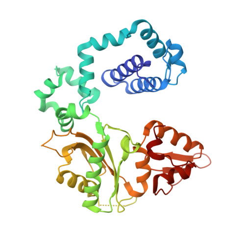 | |
UniProt & NIH Common Fund Data Resources | |||||
PHAROS: P06746 GTEx: ENSG00000070501 | |||||
Entity Groups | |||||
| Sequence Clusters | 30% Identity50% Identity70% Identity90% Identity95% Identity100% Identity | ||||
| UniProt Group | P06746 | ||||
Sequence AnnotationsExpand | |||||
Reference Sequence | |||||
Entity ID: 2 | ||||
| Molecule | Chains | Length | Organism | Image |
|---|---|---|---|---|
| DNA (5'-D(*CP*CP*GP*AP*CP*TP*GP*CP*GP*CP*AP*TP*CP*AP*GP*C)-3') | B [auth T] | 16 | synthetic construct | 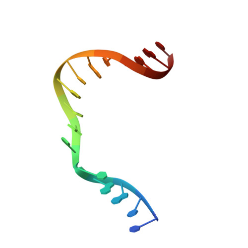 |
Sequence AnnotationsExpand | ||||
Reference Sequence | ||||
Entity ID: 3 | ||||
| Molecule | Chains | Length | Organism | Image |
|---|---|---|---|---|
| DNA (5'-D(*GP*CP*TP*GP*AP*TP*GP*CP*GP*C)-3') | C [auth P] | 10 | synthetic construct | 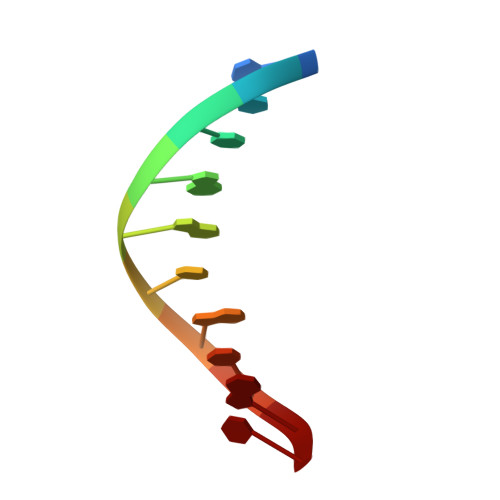 |
Sequence AnnotationsExpand | ||||
Reference Sequence | ||||
Entity ID: 4 | ||||
| Molecule | Chains | Length | Organism | Image |
|---|---|---|---|---|
| DNA (5'-D(P*GP*TP*CP*GP*G)-3') | 5 | synthetic construct | 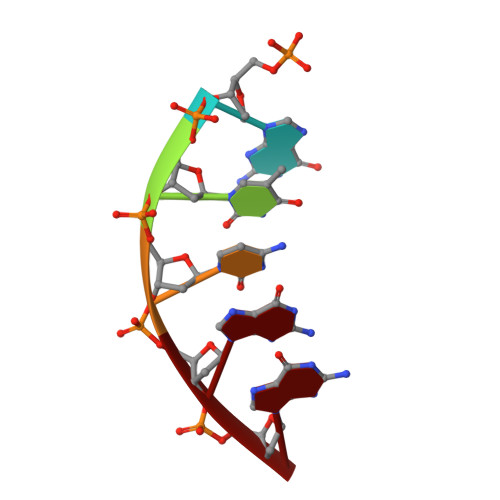 | |
Sequence AnnotationsExpand | ||||
Reference Sequence | ||||
| Ligands 3 Unique | |||||
|---|---|---|---|---|---|
| ID | Chains | Name / Formula / InChI Key | 2D Diagram | 3D Interactions | |
| XG4 Download:Ideal Coordinates CCD File | I [auth A] | 2'-deoxy-5'-O-[(R)-hydroxy{[(R)-hydroxy(phosphonooxy)phosphoryl]amino}phosphoryl]guanosine C10 H17 N6 O12 P3 DWGAAFQEGIMTIA-KVQBGUIXSA-N |  | ||
| MG Download:Ideal Coordinates CCD File | E [auth A], F [auth A] | MAGNESIUM ION Mg JLVVSXFLKOJNIY-UHFFFAOYSA-N |  | ||
| NA Download:Ideal Coordinates CCD File | G [auth A], H [auth A] | SODIUM ION Na FKNQFGJONOIPTF-UHFFFAOYSA-N |  | ||
| Length ( Å ) | Angle ( ˚ ) |
|---|---|
| a = 54.535 | α = 90 |
| b = 79.093 | β = 105.78 |
| c = 54.796 | γ = 90 |
| Software Name | Purpose |
|---|---|
| DENZO | data reduction |
| PHENIX | refinement |
| PDB_EXTRACT | data extraction |
| REFMAC | refinement |
| Funding Organization | Location | Grant Number |
|---|---|---|
| National Institutes of Health/National Institute of Environmental Health Sciences (NIH/NIEHS) | United States | ES23101 |


