4PJZ

CRYSTAL STRUCTURE OF T4 LYSOZYME-GSS-PEPTIDE IN COMPLEX WITH TEICOPLANIN-A2-2


Experimental Data Snapshot

  • Method: X-RAY DIFFRACTION
  • Resolution: 1.87 Å
  • R-Value Free: 
    0.228 (Depositor), 0.230 (DCC) 
  • R-Value Work: 
    0.194 (Depositor), 0.200 (DCC) 
  • R-Value Observed: 
    0.196 (Depositor) 

wwPDB Validation   3D Report Full Report


Ligand Structure Quality Assessment 

Created with Raphaël 2.3.0Worse 01 BetterLigand structure goodness of fit to experimental dataBest fitted NAGClick on this verticalbar to view detailsBest fitted MANClick on this verticalbar to view detailsBest fitted GCSClick on this verticalbar to view detailsBest fitted T55Click on this verticalbar to view details

This is version 2.2 of the entry. See complete history


Literature

X-ray Crystal Structure of Teicoplanin A2-2 Bound to a Catalytic Peptide Sequence via the Carrier Protein Strategy.

Han, S.Le, B.V.Hajare, H.S.Baxter, R.H.Miller, S.J.

(2014) J Org Chem 79: 8550-8556

  • DOI: https://doi.org/10.1021/jo501625f
  • Primary Citation of Related Structures:  
    4PJZ, 4PK0

  • PubMed Abstract: 

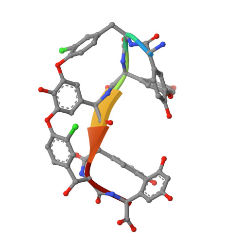
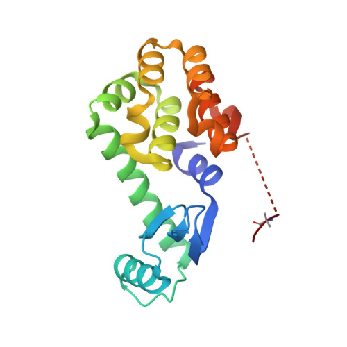
    We report the X-ray crystal structure of a site-selective peptide catalyst moiety and teicoplanin A2-2 complex. The expressed protein ligation technique was used to couple T4 lysozyme (T4L) and a synthetic peptide catalyst responsible for the selective phosphorylation of the N-acetylglucosamine sugar in a teicoplanin A2-2 derivative. The T4L-Pmh-dPro-Aib-dAla-dAla construct was crystallized in the presence of teicoplanin A2-2. The resulting 2.3 Å resolution protein-peptide-teicoplanin complex crystal structure revealed that the nucleophilic nitrogen of N-methylimidazole in the Pmh residue is in closer proximity (7.6 Å) to the N-acetylglucosamine than the two other sugar rings present in teicoplanin (9.3 and 20.3 Å, respectively). This molecular arrangement is consistent with the observed selectivity afforded by the peptide-based catalyst when it is applied to a site-selective phosphorylation reaction involving a teicoplanin A2-2 derivative.


  • Organizational Affiliation

    Department of Chemistry, Yale University , New Haven, Connecticut 06511, United States.


Macromolecules

Find similar proteins by:  Sequence   |   3D Structure  

Entity ID: 1
MoleculeChains Sequence LengthOrganismDetailsImage
TEICOPLANIN-A2-2A [auth B]7Actinoplanes teichomyceticusMutation(s): 0 
Sequence Annotations
Expand
  • Reference Sequence
Find similar proteins by:  (by identity cutoff)  |  3D Structure
Entity ID: 2
MoleculeChains Sequence LengthOrganismDetailsImage
LysozymeB [auth A]174Tequatrovirus T4Mutation(s): 2 
Gene Names: eT4Tp126
EC: 3.2.1.17
UniProt
Find proteins for D9IEF7 (Enterobacteria phage T4)
Explore D9IEF7 
Go to UniProtKB:  D9IEF7
Entity Groups  
Sequence Clusters30% Identity50% Identity70% Identity90% Identity95% Identity100% Identity
UniProt GroupD9IEF7
Sequence Annotations
Expand
  • Reference Sequence
Small Molecules
Ligands 6 Unique
IDChains Name / Formula / InChI Key2D Diagram3D Interactions
NAG
Query on NAG

Download Ideal Coordinates CCD File 
E [auth B]2-acetamido-2-deoxy-beta-D-glucopyranose
C8 H15 N O6
OVRNDRQMDRJTHS-FMDGEEDCSA-N
MAN
Query on MAN

Download Ideal Coordinates CCD File 
F [auth B]alpha-D-mannopyranose
C6 H12 O6
WQZGKKKJIJFFOK-PQMKYFCFSA-N
GCS
Query on GCS

Download Ideal Coordinates CCD File 
C [auth B]2-amino-2-deoxy-beta-D-glucopyranose
C6 H13 N O5
MSWZFWKMSRAUBD-QZABAPFNSA-N
T55
Query on T55

Download Ideal Coordinates CCD File 
D [auth B]8-METHYLNONANOIC ACID
C10 H20 O2
OAOABCKPVCUNKO-UHFFFAOYSA-N
PO4
Query on PO4

Download Ideal Coordinates CCD File 
G [auth B],
I [auth A]
PHOSPHATE ION
O4 P
NBIIXXVUZAFLBC-UHFFFAOYSA-K
GOL
Query on GOL

Download Ideal Coordinates CCD File 
H [auth B],
J [auth A]
GLYCEROL
C3 H8 O3
PEDCQBHIVMGVHV-UHFFFAOYSA-N
Modified Residues  5 Unique
IDChains TypeFormula2D DiagramParent
3MY
Query on 3MY
A [auth B]D-PEPTIDE LINKINGC9 H10 Cl N O3TYR
OMY
Query on OMY
A [auth B]L-PEPTIDE LINKINGC9 H10 Cl N O4TYR
AIB
Query on AIB
B [auth A]L-PEPTIDE LINKINGC4 H9 N O2ALA
CCS
Query on CCS
B [auth A]L-PEPTIDE LINKINGC5 H9 N O4 SCYS
MHS
Query on MHS
B [auth A]L-PEPTIDE LINKINGC7 H11 N3 O2HIS
Biologically Interesting Molecules (External Reference) 1 Unique
Experimental Data & Validation

Experimental Data

  • Method: X-RAY DIFFRACTION
  • Resolution: 1.87 Å
  • R-Value Free:  0.228 (Depositor), 0.230 (DCC) 
  • R-Value Work:  0.194 (Depositor), 0.200 (DCC) 
  • R-Value Observed: 0.196 (Depositor) 
Space Group: I 2 2 2
Unit Cell:
Length ( Å )Angle ( ˚ )
a = 58.116α = 90
b = 63.569β = 90
c = 138.884γ = 90
Software Package:
Software NamePurpose
REFMACrefinement

Structure Validation

View Full Validation Report


Ligand Structure Quality Assessment 

Created with Raphaël 2.3.0Worse 01 BetterLigand structure goodness of fit to experimental dataBest fitted NAGClick on this verticalbar to view detailsBest fitted MANClick on this verticalbar to view detailsBest fitted GCSClick on this verticalbar to view detailsBest fitted T55Click on this verticalbar to view details

Entry History & Funding Information

Deposition Data


Funding OrganizationLocationGrant Number
National Institutes of Health/National Institute of General Medical Sciences (NIH/NIGMS)United StatesGM-068649

Revision History  (Full details and data files)

  • Version 1.0: 2014-09-10
    Type: Initial release
  • Version 1.1: 2014-10-01
    Changes: Database references
  • Version 1.2: 2017-09-20
    Changes: Author supporting evidence, Database references, Derived calculations, Other, Source and taxonomy, Structure summary
  • Version 2.0: 2019-12-25
    Changes: Author supporting evidence, Polymer sequence
  • Version 2.1: 2020-07-29
    Type: Remediation
    Reason: Carbohydrate remediation
    Changes: Data collection, Derived calculations, Refinement description, Structure summary
  • Version 2.2: 2023-12-27
    Changes: Data collection, Database references, Derived calculations, Structure summary