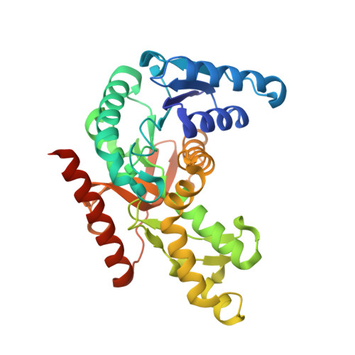
4PLT

Crystal structure of ancestral apicomplexan malate dehydrogenase with oxamate.


Experimental Data Snapshot

  • Method: X-RAY DIFFRACTION
  • Resolution: 1.60 Å
  • R-Value Free: 0.221 
  • R-Value Work: 0.189 
  • R-Value Observed: 0.190 

Starting Model: experimental
View more details

wwPDB Validation   3D Report Full Report


Ligand Structure Quality Assessment 


This is version 1.8 of the entry. See complete history


Literature

An atomic-resolution view of neofunctionalization in the evolution of apicomplexan lactate dehydrogenases.

Boucher, J.I.Jacobowitz, J.R.Beckett, B.C.Classen, S.Theobald, D.L.

(2014) Elife 3: e02304-e02304

  • DOI: https://doi.org/10.7554/eLife.02304
  • Primary Citation of Related Structures:  
    4PLC, 4PLF, 4PLG, 4PLH, 4PLT, 4PLV, 4PLW, 4PLY, 4PLZ

  • PubMed Abstract: 

    Malate and lactate dehydrogenases (MDH and LDH) are homologous, core metabolic enzymes that share a fold and catalytic mechanism yet possess strict specificity for their substrates. In the Apicomplexa, convergent evolution of an unusual LDH from MDH produced a difference in specificity exceeding 12 orders of magnitude. The mechanisms responsible for this extraordinary functional shift are currently unknown. Using ancestral protein resurrection, we find that specificity evolved in apicomplexan LDHs by classic neofunctionalization characterized by long-range epistasis, a promiscuous intermediate, and few gain-of-function mutations of large effect. In canonical MDHs and LDHs, a single residue in the active-site loop governs substrate specificity: Arg102 in MDHs and Gln102 in LDHs. During the evolution of the apicomplexan LDH, however, specificity switched via an insertion that shifted the position and identity of this 'specificity residue' to Trp107f. Residues far from the active site also determine specificity, as shown by the crystal structures of three ancestral proteins bracketing the key duplication event. This work provides an unprecedented atomic-resolution view of evolutionary trajectories creating a nascent enzymatic function.


  • Organizational Affiliation

    Department of Biochemistry, Brandeis University, Waltham, United States.


Macromolecules
Find similar proteins by:  (by identity cutoff)  |  3D Structure
Entity ID: 1
MoleculeChains Sequence LengthOrganismDetailsImage
malate dehydrogenaseA [auth B],
B [auth A],
C,
D
331ApicomplexaMutation(s): 0 
UniProt
Find proteins for A0A075B5H0 (Apicomplexa)
Explore A0A075B5H0 
Go to UniProtKB:  A0A075B5H0
Entity Groups  
Sequence Clusters30% Identity50% Identity70% Identity90% Identity95% Identity100% Identity
UniProt GroupA0A075B5H0
Sequence Annotations
Expand
  • Reference Sequence
Small Molecules
Ligands 2 Unique
IDChains Name / Formula / InChI Key2D Diagram3D Interactions
NAI
Query on NAI

Download Ideal Coordinates CCD File 
E [auth B],
G [auth A],
I [auth C],
K [auth D]
1,4-DIHYDRONICOTINAMIDE ADENINE DINUCLEOTIDE
C21 H29 N7 O14 P2
BOPGDPNILDQYTO-NNYOXOHSSA-N
OXM
Query on OXM

Download Ideal Coordinates CCD File 
F [auth B],
H [auth A],
J [auth C],
L [auth D]
OXAMIC ACID
C2 H3 N O3
SOWBFZRMHSNYGE-UHFFFAOYSA-N
Experimental Data & Validation

Experimental Data

  • Method: X-RAY DIFFRACTION
  • Resolution: 1.60 Å
  • R-Value Free: 0.221 
  • R-Value Work: 0.189 
  • R-Value Observed: 0.190 
  • Space Group: P 1 21 1
Unit Cell:
Length ( Å )Angle ( ˚ )
a = 59.527α = 90
b = 136.311β = 108.78
c = 90.238γ = 90
Software Package:
Software NamePurpose
Blu-Icedata collection
XDSdata reduction
Cootmodel building
PHENIXrefinement
PDB_EXTRACTdata extraction

Structure Validation

View Full Validation Report


Ligand Structure Quality Assessment 


Entry History & Funding Information

Deposition Data


Funding OrganizationLocationGrant Number
National Institutes of Health/National Institute of General Medical Sciences (NIH/NIGMS)United StatesR01GM096053
National Institutes of Health/National Institute of General Medical Sciences (NIH/NIGMS)United StatesR01GM094468
National Institutes of Health/National Institute of General Medical Sciences (NIH/NIGMS)United StatesR01GM105404

Revision History  (Full details and data files)

  • Version 1.0: 2014-07-02
    Type: Initial release
  • Version 1.1: 2014-07-16
    Changes: Database references
  • Version 1.2: 2014-09-24
    Changes: Structure summary
  • Version 1.3: 2014-10-01
    Changes: Database references
  • Version 1.4: 2014-12-24
    Changes: Database references
  • Version 1.5: 2015-02-25
    Changes: Derived calculations
  • Version 1.6: 2017-09-27
    Changes: Author supporting evidence, Derived calculations, Other, Source and taxonomy, Structure summary
  • Version 1.7: 2019-12-25
    Changes: Author supporting evidence
  • Version 1.8: 2023-09-27
    Changes: Data collection, Database references, Refinement description