Discovery of new acylaminopyridines as GSK-3 inhibitors by a structure guided in-depth exploration of chemical space around a pyrrolopyridinone core.
Sivaprakasam, P., Han, X., Civiello, R.L., Jacutin-Porte, S., Kish, K., Pokross, M., Lewis, H.A., Ahmed, N., Szapiel, N., Newitt, J.A., Baldwin, E.T., Xiao, H., Krause, C.M., Park, H., Nophsker, M., Lippy, J.S., Burton, C.R., Langley, D.R., Macor, J.E., Dubowchik, G.M.(2015) Bioorg Med Chem Lett 25: 1856-1863
- PubMed: 25845281 Search on PubMed
- DOI: https://doi.org/10.1016/j.bmcl.2015.03.046
- Primary Citation Related Structures:
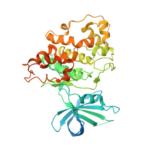
4PTC, 4PTE, 4PTG - PubMed Abstract:
Glycogen synthase kinase-3 (GSK-3) has been proposed to play a crucial role in the pathogenesis of many diseases including cancer, stroke, bipolar disorders, diabetes and neurodegenerative diseases. GSK-3 inhibition has been a major area of pharmaceutical interest over the last two decades. A plethora of reports appeared recently on selective inhibitors and their co-crystal structures in GSK-3β. We identified several series of promising new GSK-3β inhibitors from a coherent design around a pyrrolopyridinone core structure. A systematic exploration of the chemical space around the central spacer led to potent single digit and sub-nanomolar GSK-3β inhibitors. When dosed orally in a transgenic mouse model of Alzheimer's disease (AD), an exemplary compound showed significant lowering of Tau phosphorylation at one of the GSK-3 phosphorylating sites, Ser396. X-ray crystallography greatly aided in validating the binding hypotheses.
- Bristol-Myers Squibb Research and Development, 5 Research Parkway, Wallingford, CT 06492, USA. Electronic address: prasanna.siva@bms.com.
Organizational Affiliation: