


Aldose Reductase: How expensive is the opening of the specificity pocket? IDD ligands under investigation
Rechlin, C., Heine, A., Scheer, F., Toth, P., Diederich, W., Klebe, G.To be published.
Experimental Data Snapshot
Starting Model: experimental
View more details
Entity ID: 1 | |||||
|---|---|---|---|---|---|
| Molecule | Chains | Sequence Length | Organism | Details | Image |
| Aldose reductase | 316 | Homo sapiens | Mutation(s): 0 Gene Names: AKR1B1, ALDR1 EC: 1.1.1.21 (PDB Primary Data), 1.1.1.372 (UniProt), 1.1.1.300 (UniProt), 1.1.1.54 (UniProt) | 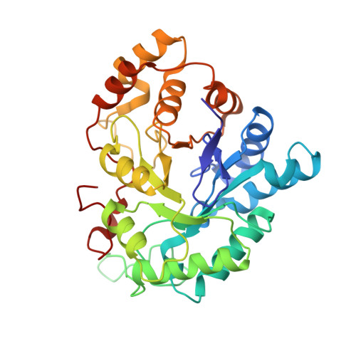 | |
UniProt & NIH Common Fund Data Resources | |||||
GTEx: ENSG00000085662 | |||||
Entity Groups | |||||
| Sequence Clusters | 30% Identity50% Identity70% Identity90% Identity95% Identity100% Identity | ||||
| UniProt Group | P15121 | ||||
Sequence AnnotationsExpand | |||||
Reference Sequence | |||||
| Ligands 3 Unique | |||||
|---|---|---|---|---|---|
| ID | Chains | Name / Formula / InChI Key | 2D Diagram | 3D Interactions | |
| NAP Download:Ideal Coordinates CCD File | D [auth A] | NADP NICOTINAMIDE-ADENINE-DINUCLEOTIDE PHOSPHATE C21 H28 N7 O17 P3 XJLXINKUBYWONI-NNYOXOHSSA-N |  | ||
| 2WQ Download:Ideal Coordinates CCD File | B [auth A] | [5-fluoro-2-(prop-2-yn-1-ylcarbamoyl)phenoxy]acetic acid C12 H10 F N O4 PZTOBGBKDQCKIG-UHFFFAOYSA-N |  | ||
| CIT Download:Ideal Coordinates CCD File | C [auth A] | CITRIC ACID C6 H8 O7 KRKNYBCHXYNGOX-UHFFFAOYSA-N |  | ||
| Length ( Å ) | Angle ( ˚ ) |
|---|---|
| a = 49.411 | α = 90 |
| b = 66.688 | β = 92.02 |
| c = 47.353 | γ = 90 |
| Software Name | Purpose |
|---|---|
| MAR345dtb | data collection |
| PHASER | phasing |
| PHENIX | refinement |
| HKL-2000 | data reduction |
| HKL-2000 | data scaling |


