4QY3 | pdb_00004qy3

THE CRYSTAL STRUCTURE OF THE COMPLEX of HCAII WITH AN ORTHO-SUBSTITUTED BENZOIC ACID


Experimental Data Snapshot

  • Method: X-RAY DIFFRACTION
  • Resolution: 1.50 Å
  • R-Value Free: 
    0.184 (Depositor), 0.178 (DCC) 
  • R-Value Work: 
    0.163 (Depositor), 0.159 (DCC) 

Starting Model: experimental
View more details

wwPDB Validation 3D Report Full Report

Validation slider image for 4QY3

Ligand Structure Quality Assessment 


This is version 1.2 of the entry. See complete history

Literature

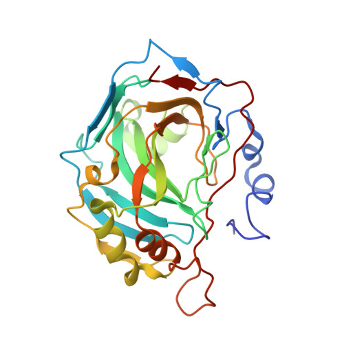
Out of the active site binding pocket for carbonic anhydrase inhibitors.

D'Ambrosio, K.Carradori, S.Monti, S.M.Buonanno, M.Secci, D.Vullo, D.Supuran, C.T.De Simone, G.

(2014) Chem Commun (Camb) 51: 302-305

  • DOI: https://doi.org/10.1039/c4cc07320g
  • Primary Citation Related Structures: 
    4QY3

  • PubMed Abstract: 

    A structural study of the adduct which 2-benzylsulfinylbenzoic acid forms with human carbonic anhydrase II is reported, showing a binding mode completely different from any other class of carbonic anhydrase inhibitors investigated so far; this carboxylate binds in a pocket situated out of the enzyme active site.


  • Organizational Affiliation
    • Istituto di Biostrutture e Bioimmagini-CNR, Via Mezzocannone 16, 80134, Naples, Italy. gdesimon@unina.it katia.dambrosio@cnr.it.

Macromolecule Content 

  • Total Structure Weight: 29.82 kDa 
  • Atom Count: 2,414 
  • Modeled Residue Count: 257 
  • Deposited Residue Count: 259 
  • Unique protein chains: 1

Macromolecules

Find similar proteins by:|  3D Structure
Entity ID: 1
MoleculeChains  Sequence LengthOrganismDetailsImage
Carbonic anhydrase 2259Homo sapiensMutation(s): 0 
Gene Names: CA2
EC: 4.2.1.1 (PDB Primary Data), 4.2.1.69 (UniProt)
UniProt & NIH Common Fund Data Resources
Find proteins for P00918 (Homo sapiens)
Explore P00918 
Go to UniProtKB:  P00918
PHAROS:  P00918
GTEx:  ENSG00000104267 
Entity Groups
Sequence Clusters30% Identity50% Identity70% Identity90% Identity95% Identity100% Identity
UniProt GroupP00918
Sequence Annotations
Expand
Reference Sequence

Experimental Data & Validation

Experimental Data

  • Method: X-RAY DIFFRACTION
  • Resolution: 1.50 Å
  • R-Value Free:  0.184 (Depositor), 0.178 (DCC) 
  • R-Value Work:  0.163 (Depositor), 0.159 (DCC) 
Space Group: P 1 21 1
Unit Cell:
Length ( Å )Angle ( ˚ )
a = 42.15α = 90
b = 41.53β = 104.52
c = 72.18γ = 90
Software Package:
Software NamePurpose
CNSrefinement
HKL-2000data reduction
HKL-2000data scaling
CNSphasing

Structure Validation

View Full Validation Report


Ligand Structure Quality Assessment 


Entry History 

Deposition Data

Revision History  (Full details and data files)

  • Version 1.0: 2014-12-03
    Type: Initial release
  • Version 1.1: 2014-12-17
    Changes: Database references
  • Version 1.2: 2023-09-20
    Changes: Data collection, Database references, Derived calculations, Refinement description