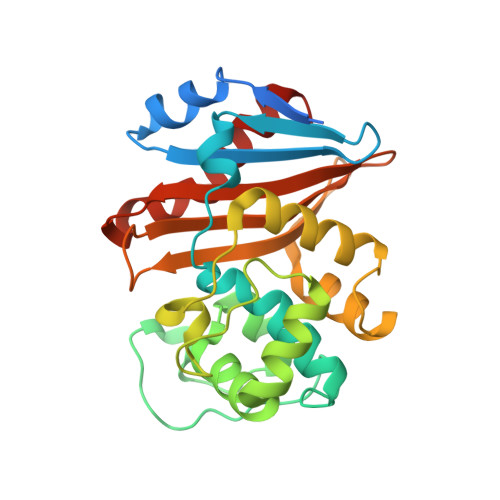
4S2K

OXA-48 in complex with Avibactam at pH 7.5


Experimental Data Snapshot

  • Method: X-RAY DIFFRACTION
  • Resolution: 2.10 Å
  • R-Value Free: 0.206 
  • R-Value Work: 0.175 
  • R-Value Observed: 0.177 

Starting Model: experimental
View more details

wwPDB Validation   3D Report Full Report


Ligand Structure Quality Assessment 


This is version 1.5 of the entry. See complete history


Literature

Molecular Mechanism of Avibactam-Mediated beta-Lactamase Inhibition.

King, D.T.King, A.M.Lal, S.M.Wright, G.D.Strynadka, N.C.

(2015) ACS Infect Dis 1: 175-184

  • DOI: https://doi.org/10.1021/acsinfecdis.5b00007
  • Primary Citation of Related Structures:  
    4S2I, 4S2J, 4S2K, 4S2N, 4S2O, 4S2P

  • PubMed Abstract: 

    Emerging β-lactamase-mediated resistance is threatening the clinical utility of the single most prominent class of antibacterial agents used in medicine, the β-lactams. The diazabicyclooctane avibactam is able to inhibit a wider range of serine β-lactamases than has been previously observed with β-lactamase inhibitors such as the widely prescribed clavulanic acid. However, despite its broad-spectrum activity, variable levels of inhibition have been observed for molecular class D β-lactamases. In order to better understand the molecular basis and spectrum of inhibition by avibactam, we provide structural and mechanistic analysis of the compound in complex with important class A and D serine β-lactamases. Herein, we reveal the 1.7- and 2.0-Å-resolution crystal structures of avibactam covalently bound to class D β-lactamases OXA-10 and OXA-48. Furthermore, a kinetic analysis of key active-site mutants for class A β-lactamase CTX-M-15 allows us to propose a validated mechanism for avibactam-mediated β-lactamase inhibition including a unique role for S130, which acts as a general base. This study provides molecular insights that will aid in the design and development of avibactam-based chemotherapeutic agents effective against emerging drug-resistant microorganisms.


  • Organizational Affiliation

    The Department of Biochemistry and Molecular Biology and Center for Blood Research, University of British Columbia , 2350 Health Sciences Mall, Vancouver, British Columbia V6T 1Z3, Canada.


Macromolecules
Find similar proteins by:  (by identity cutoff)  |  3D Structure
Entity ID: 1
MoleculeChains Sequence LengthOrganismDetailsImage
Beta-lactamase
A, B, C, D
265Klebsiella pneumoniaeMutation(s): 0 
Gene Names: bla OXA-48blaOXA-48FP68_27275KPE71T_00045
EC: 3.5.2.6
UniProt
Find proteins for Q6XEC0 (Klebsiella pneumoniae)
Explore Q6XEC0 
Go to UniProtKB:  Q6XEC0
Entity Groups  
Sequence Clusters30% Identity50% Identity70% Identity90% Identity95% Identity100% Identity
UniProt GroupQ6XEC0
Sequence Annotations
Expand
  • Reference Sequence
Experimental Data & Validation

Experimental Data

  • Method: X-RAY DIFFRACTION
  • Resolution: 2.10 Å
  • R-Value Free: 0.206 
  • R-Value Work: 0.175 
  • R-Value Observed: 0.177 
  • Space Group: P 32
Unit Cell:
Length ( Å )Angle ( ˚ )
a = 142.78α = 90
b = 142.78β = 90
c = 52.42γ = 120
Software Package:
Software NamePurpose
MxDCdata collection
PHASERphasing
BUSTERrefinement
MOSFLMdata reduction
SCALEPACKdata scaling

Structure Validation

View Full Validation Report


Ligand Structure Quality Assessment 


Entry History 

Deposition Data

Revision History  (Full details and data files)

  • Version 1.0: 2015-02-25
    Type: Initial release
  • Version 1.1: 2015-06-03
    Changes: Non-polymer description
  • Version 1.2: 2016-09-28
    Changes: Database references
  • Version 1.3: 2017-10-25
    Changes: Author supporting evidence, Refinement description
  • Version 1.4: 2023-09-20
    Changes: Data collection, Database references, Derived calculations, Refinement description, Structure summary
  • Version 1.5: 2024-10-30
    Changes: Structure summary