4U2U

Bak domain swapped dimer induced by BidBH3 with CHAPS


Experimental Data Snapshot

  • Method: X-RAY DIFFRACTION
  • Resolution: 2.90 Å
  • R-Value Free: 0.263 
  • R-Value Work: 0.209 
  • R-Value Observed: 0.214 

wwPDB Validation   3D Report Full Report


This is version 1.4 of the entry. See complete history


Literature

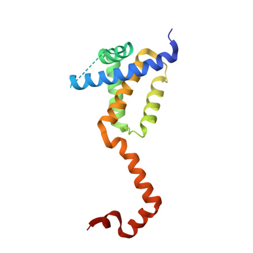
Bak Core and Latch Domains Separate during Activation, and Freed Core Domains Form Symmetric Homodimers.

Brouwer, J.M.Westphal, D.Dewson, G.Robin, A.Y.Uren, R.T.Bartolo, R.Thompson, G.V.Colman, P.M.Kluck, R.M.Czabotar, P.E.

(2014) Mol Cell 55: 938-946

  • DOI: https://doi.org/10.1016/j.molcel.2014.07.016
  • Primary Citation of Related Structures:  
    4U2U, 4U2V

  • PubMed Abstract: 

    Apoptotic stimuli activate and oligomerize the proapoptotic proteins Bak and Bax, resulting in mitochondrial outer-membrane permeabilization and subsequent cell death. This activation can occur when certain BH3-only proteins interact directly with Bak and Bax. Recently published crystal structures reveal that Bax separates into core and latch domains in response to BH3 peptides. The distinguishing characteristics of BH3 peptides capable of directly activating Bax were also elucidated. Here we identify specific BH3 peptides capable of "unlatching" Bak and describe structural insights into Bak activation and oligomerization. Crystal structures and crosslinking experiments demonstrate that Bak undergoes a conformational change similar to that of Bax upon activation. A structure of the Bak core domain dimer provides a high-resolution image of this key intermediate in the pore-forming oligomer. Our results confirm an analogous mechanism for activation and dimerization of Bak and Bax in response to certain BH3 peptides.


  • Organizational Affiliation

    Walter and Eliza Hall Institute of Medical Research, 1G Royal Parade, Parkville, Victoria 3052, Australia; Department of Medical Biology, The University of Melbourne, Melbourne, Victoria 3052, Australia.


Macromolecules
Find similar proteins by:  (by identity cutoff)  |  3D Structure
Entity ID: 1
MoleculeChains Sequence LengthOrganismDetailsImage
Bcl-2 homologous antagonist/killer
A, B
169Homo sapiensMutation(s): 1 
Gene Names: BAK1BAKBCL2L7CDN1
UniProt & NIH Common Fund Data Resources
Find proteins for Q16611 (Homo sapiens)
Explore Q16611 
Go to UniProtKB:  Q16611
PHAROS:  Q16611
GTEx:  ENSG00000030110 
Entity Groups  
Sequence Clusters30% Identity50% Identity70% Identity90% Identity95% Identity100% Identity
UniProt GroupQ16611
Sequence Annotations
Expand
  • Reference Sequence
Experimental Data & Validation

Experimental Data

  • Method: X-RAY DIFFRACTION
  • Resolution: 2.90 Å
  • R-Value Free: 0.263 
  • R-Value Work: 0.209 
  • R-Value Observed: 0.214 
  • Space Group: C 1 2 1
Unit Cell:
Length ( Å )Angle ( ˚ )
a = 110.646α = 90
b = 39.482β = 117.71
c = 88.147γ = 90
Software Package:
Software NamePurpose
XDSdata reduction
XSCALEdata scaling
BUSTER-TNTrefinement
PDB_EXTRACTdata extraction
PHASERphasing
BUSTERrefinement

Structure Validation

View Full Validation Report


Entry History & Funding Information

Deposition Data


Funding OrganizationLocationGrant Number
National Health and Medical Research Council (NHMRC, Australia)Australia1059331
National Health and Medical Research Council (NHMRC, Australia)Australia1023055
National Health and Medical Research Council (NHMRC, Australia)Australia1016701

Revision History  (Full details and data files)

  • Version 1.0: 2014-09-10
    Type: Initial release
  • Version 1.1: 2014-10-01
    Changes: Database references
  • Version 1.2: 2017-09-27
    Changes: Author supporting evidence, Data collection, Database references, Derived calculations, Other, Source and taxonomy
  • Version 1.3: 2020-01-08
    Changes: Author supporting evidence
  • Version 1.4: 2023-09-27
    Changes: Data collection, Database references, Refinement description