4UE1

Structure of the stapled peptide YS-01 bound to MDM2


Experimental Data Snapshot

  • Method: X-RAY DIFFRACTION
  • Resolution: 1.45 Å
  • R-Value Free: 0.184 
  • R-Value Work: 0.140 
  • R-Value Observed: 0.143 

wwPDB Validation   3D Report Full Report


This is version 2.1 of the entry. See complete history


Literature

Benzene Probes in Molecular Dynamics Simulations Reveal Novel Binding Sites for Ligand Design.

Tan, Y.S.Reeks, J.Brown, C.J.Thean, D.Ferrer Gago, F.J.Yuen, T.Y.Goh, E.T.Lee, X.E.Jennings, C.E.Joseph, T.L.Lakshminarayanan, R.Lane, D.P.Noble, M.E.Verma, C.S.

(2016) J Phys Chem Lett 7: 3452-3457

  • DOI: https://doi.org/10.1021/acs.jpclett.6b01525
  • Primary Citation of Related Structures:  
    4UD7, 4UE1

  • PubMed Abstract: 

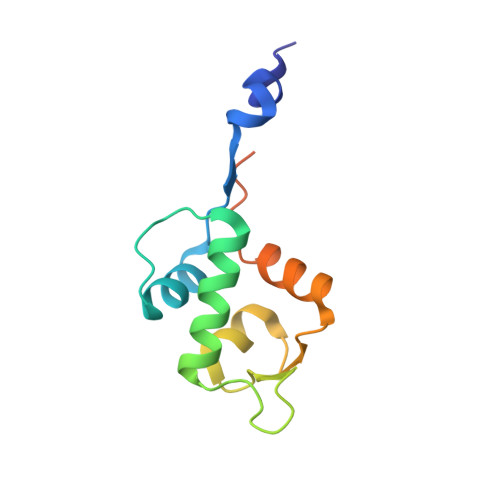
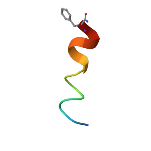
    Protein flexibility poses a major challenge in binding site identification. Several computational pocket detection methods that utilize small-molecule probes in molecular dynamics (MD) simulations have been developed to address this issue. Although they have proven hugely successful at reproducing experimental structural data, their ability to predict new binding sites that are yet to be identified and characterized has not been demonstrated. Here, we report the use of benzenes as probe molecules in ligand-mapping MD (LMMD) simulations to predict the existence of two novel binding sites on the surface of the oncoprotein MDM2. One of them was serendipitously confirmed by biophysical assays and X-ray crystallography to be important for the binding of a new family of hydrocarbon stapled peptides that were specifically designed to target the other putative site. These results highlight the predictive power of LMMD and suggest that predictions derived from LMMD simulations can serve as a reliable basis for the identification of novel ligand binding sites in structure-based drug design.


  • Organizational Affiliation

    Bioinformatics Institute, Agency for Science, Technology and Research (A*STAR) , 30 Biopolis Street, #07-01 Matrix, Singapore 138671.


Macromolecules
Find similar proteins by:  (by identity cutoff)  |  3D Structure
Entity ID: 1
MoleculeChains Sequence LengthOrganismDetailsImage
E3 UBIQUITIN-PROTEIN LIGASE MDM2
A, B, C, D
114Homo sapiensMutation(s): 2 
EC: 6.3.2
UniProt & NIH Common Fund Data Resources
Find proteins for Q00987 (Homo sapiens)
Explore Q00987 
Go to UniProtKB:  Q00987
PHAROS:  Q00987
GTEx:  ENSG00000135679 
Entity Groups  
Sequence Clusters30% Identity50% Identity70% Identity90% Identity95% Identity100% Identity
UniProt GroupQ00987
Sequence Annotations
Expand
  • Reference Sequence

Find similar proteins by:  Sequence   |   3D Structure  

Entity ID: 2
MoleculeChains Sequence LengthOrganismDetailsImage
YS-01E [auth F],
F [auth G],
G [auth H],
H [auth I]
16Homo sapiensMutation(s): 0 
Entity Groups  
Sequence Clusters30% Identity50% Identity70% Identity90% Identity95% Identity100% Identity
Sequence Annotations
Expand
  • Reference Sequence
Experimental Data & Validation

Experimental Data

  • Method: X-RAY DIFFRACTION
  • Resolution: 1.45 Å
  • R-Value Free: 0.184 
  • R-Value Work: 0.140 
  • R-Value Observed: 0.143 
  • Space Group: P 1 21 1
Unit Cell:
Length ( Å )Angle ( ˚ )
a = 46.104α = 90
b = 69.462β = 101.96
c = 78.562γ = 90
Software Package:
Software NamePurpose
REFMACrefinement
XDSdata reduction
Aimlessdata scaling
PHASERphasing

Structure Validation

View Full Validation Report


Entry History 

Deposition Data

Revision History  (Full details and data files)

  • Version 1.0: 2016-01-13
    Type: Initial release
  • Version 1.1: 2017-01-18
    Changes: Database references
  • Version 1.2: 2023-09-27
    Changes: Data collection, Database references, Derived calculations, Other
  • Version 2.0: 2023-11-15
    Changes: Atomic model, Data collection, Derived calculations
  • Version 2.1: 2023-12-20
    Changes: Refinement description