4UMN

Structure of a stapled peptide antagonist bound to Nutlin-resistant Mdm2.


Experimental Data Snapshot

  • Method: X-RAY DIFFRACTION
  • Resolution: 1.99 Å
  • R-Value Free: 0.237 
  • R-Value Work: 0.200 
  • R-Value Observed: 0.202 

wwPDB Validation   3D Report Full Report


This is version 3.1 of the entry. See complete history


Literature

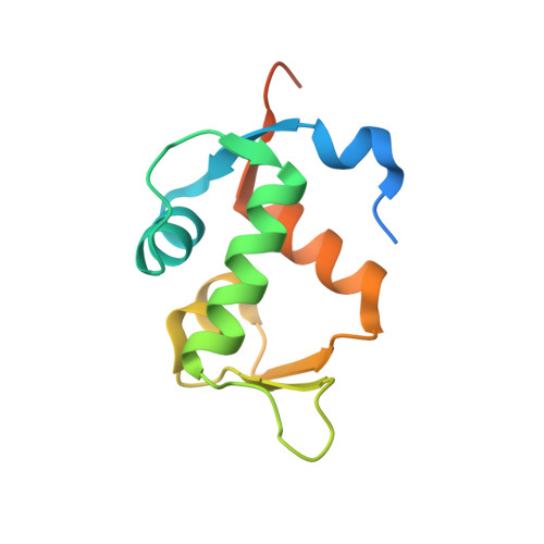
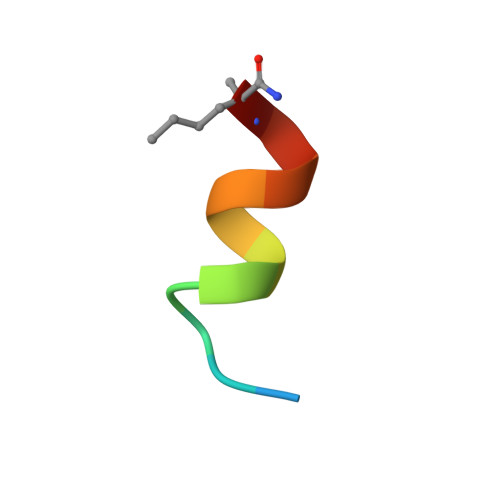
Structure of a stapled peptide antagonist bound to nutlin-resistant Mdm2.

Chee, S.M.Wongsantichon, J.Soo Tng, Q.Robinson, R.Joseph, T.L.Verma, C.Lane, D.P.Brown, C.J.Ghadessy, F.J.

(2014) PLoS One 9: e104914-e104914

  • DOI: https://doi.org/10.1371/journal.pone.0104914
  • Primary Citation of Related Structures:  
    4UMN

  • PubMed Abstract: 

    As key negative regulator of the p53 tumour suppressor, Mdm2 is an attractive therapeutic target. Small molecules such as Nutlin have been developed to antagonise Mdm2, resulting in p53-dependent death of tumour cells. We have recently described a mutation in Mdm2 (M62A), which precludes binding of Nutlin, but not p53. This Nutlin-resistant variant is not, however, refractory to binding and inhibition by stapled peptide antagonists targeting the same region of Mdm2. A detailed understanding of how stapled peptides are recalcitrant to Mdm2 mutations conferring Nutlin-resistance will aid in the further development of potent Mdm2 antagonists. Here, we report the 2.00 Å crystal structure of a stapled peptide antagonist bound to Nutlin resistant Mdm2. The stapled peptide relies on an extended network of interactions along the hydrophobic binding cleft of Mdm2 for high affinity binding. Additionally, as seen in other stapled peptide structures, the hydrocarbon staple itself contributes to binding through favourable interactions with Mdm2. The structure highlights the intrinsic plasticity present in both Mdm2 and the hydrocarbon staple moiety, and can be used to guide future iterations of both small molecules and stapled peptides for improved antagonists of Mdm2.


  • Organizational Affiliation

    p53Lab, Agency for Science Technology and Research (A*STAR), Singapore, Singapore.


Macromolecules
Find similar proteins by:  (by identity cutoff)  |  3D Structure
Entity ID: 1
MoleculeChains Sequence LengthOrganismDetailsImage
E3 ubiquitin-protein ligase Mdm2
A, B
120Homo sapiensMutation(s): 0 
Gene Names: MDM2
EC: 2.3.2.27
UniProt & NIH Common Fund Data Resources
Find proteins for Q00987 (Homo sapiens)
Explore Q00987 
Go to UniProtKB:  Q00987
PHAROS:  Q00987
GTEx:  ENSG00000135679 
Entity Groups  
Sequence Clusters30% Identity50% Identity70% Identity90% Identity95% Identity100% Identity
UniProt GroupQ00987
Sequence Annotations
Expand
  • Reference Sequence

Find similar proteins by:  Sequence   |   3D Structure  

Entity ID: 2
MoleculeChains Sequence LengthOrganismDetailsImage
M06
C, D
13synthetic constructMutation(s): 0 
Entity Groups  
Sequence Clusters30% Identity50% Identity70% Identity90% Identity95% Identity100% Identity
Sequence Annotations
Expand
  • Reference Sequence
Small Molecules
Modified Residues  1 Unique
IDChains TypeFormula2D DiagramParent
MK8
Query on MK8
C, D
L-PEPTIDE LINKINGC7 H15 N O2LEU
Experimental Data & Validation

Experimental Data

  • Method: X-RAY DIFFRACTION
  • Resolution: 1.99 Å
  • R-Value Free: 0.237 
  • R-Value Work: 0.200 
  • R-Value Observed: 0.202 
  • Space Group: P 2 21 21
Unit Cell:
Length ( Å )Angle ( ˚ )
a = 39.076α = 90
b = 65.674β = 90
c = 105.683γ = 90
Software Package:
Software NamePurpose
REFMACrefinement
HKL-2000data reduction
HKL-2000data scaling
PHASERphasing

Structure Validation

View Full Validation Report


Entry History 

Deposition Data

Revision History  (Full details and data files)

  • Version 1.0: 2014-05-28
    Type: Initial release
  • Version 1.1: 2014-07-16
    Changes: Atomic model, Other
  • Version 1.2: 2014-08-27
    Changes: Database references
  • Version 1.3: 2017-08-09
    Changes: Database references
  • Version 1.4: 2019-10-16
    Changes: Data collection, Derived calculations, Experimental preparation, Other
  • Version 2.0: 2023-03-15
    Changes: Atomic model, Database references, Derived calculations, Non-polymer description, Polymer sequence, Source and taxonomy, Structure summary
  • Version 3.0: 2023-11-15
    Changes: Atomic model, Data collection, Derived calculations
  • Version 3.1: 2024-02-07
    Changes: Refinement description