4UQL | pdb_00004uql

High-resolution structure of a Ni-A Ni-Sox mixture of the D. fructosovorans NiFe-hydrogenase L122A mutant


Experimental Data Snapshot

  • Method: X-RAY DIFFRACTION
  • Resolution: 1.22 Å
  • R-Value Free: 
    0.152 (Depositor), 0.161 (DCC) 
  • R-Value Work: 
    0.120 (Depositor), 0.132 (DCC) 
  • R-Value Observed: 
    0.122 (Depositor) 

Starting Model: experimental
View more details

wwPDB Validation 3D Report Full Report

Validation slider image for 4UQL

Ligand Structure Quality Assessment 


This is version 1.4 of the entry. See complete history

Literature

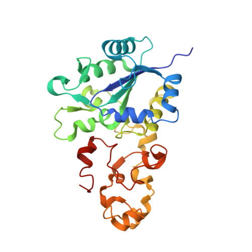
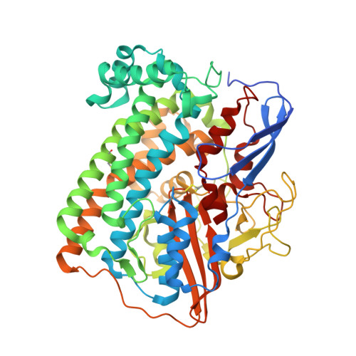
Crystallographic Studies of [Nife]-Hydrogenase Mutants: Towards Consensus Structures for the Elusive Unready Oxidized States.

Volbeda, A.Martin, L.Barbier, E.Gutierrez-Sanz, O.De Lacey, A.L.Liebgott, P.Dementin, S.Rousset, M.Fontecilla-Camps, J.C.

(2015) J Biol Inorg Chem 20: 11

  • DOI: https://doi.org/10.1007/s00775-014-1203-9
  • Primary Citation Related Structures: 
    4UPE, 4UPV, 4UQL, 4UQP, 4URH

  • PubMed Abstract: 

    Catalytically inactive oxidized O2-sensitive [NiFe]-hydrogenases are characterized by a mixture of the paramagnetic Ni-A and Ni-B states. Upon O2 exposure, enzymes in a partially reduced state preferentially form the unready Ni-A state. Because partial O2 reduction should generate a peroxide intermediate, this species was previously assigned to the elongated Ni-Fe bridging electron density observed for preparations of [NiFe]-hydrogenases known to contain the Ni-A state. However, this proposition has been challenged based on the stability of this state to UV light exposure and the possibility of generating it anaerobically under either chemical or electrochemical oxidizing conditions. Consequently, we have considered alternative structures for the Ni-A species including oxidation of thiolate ligands to either sulfenate or sulfenic acid. Here, we report both new and revised [NiFe]-hydrogenases structures and conclude, taking into account corresponding characterizations by Fourier transform infrared spectroscopy (FTIR), that the Ni-A species contains oxidized cysteine and bridging hydroxide ligands instead of the peroxide ligand we proposed earlier. Our analysis was rendered difficult by the typical formation of mixtures of unready oxidized states that, furthermore, can be reduced by X-ray induced photoelectrons. The present study could be carried out thanks to the use of Desulfovibrio fructosovorans [NiFe]-hydrogenase mutants with special properties. In addition to the Ni-A state, crystallographic results are also reported for two diamagnetic unready states, allowing the proposal of a revised oxidized inactive Ni-SU model and a new structure characterized by a persulfide ion that is assigned to an Ni-'Sox' species.


  • Organizational Affiliation
    • University Grenoble Alpes, IBS, 38044, Grenoble, France. anne.volbeda@ibs.fr.

Macromolecule Content 

  • Total Structure Weight: 183.13 kDa 
  • Atom Count: 14,688 
  • Modeled Residue Count: 1,614 
  • Deposited Residue Count: 1,656 
  • Unique protein chains: 2

Macromolecules

Find similar proteins by:|  3D Structure
Entity ID: 1
MoleculeChains  Sequence LengthOrganismDetailsImage
HYDROGENASE (NIFE) SMALL SUBUNIT HYDA
A, B
265Solidesulfovibrio fructosivoransMutation(s): 0 
EC: 1.12.2.1
UniProt
Find proteins for P18187 (Solidesulfovibrio fructosivorans)
Explore P18187 
Go to UniProtKB:  P18187
Entity Groups
Sequence Clusters30% Identity50% Identity70% Identity90% Identity95% Identity100% Identity
UniProt GroupP18187
Sequence Annotations
Expand
Reference Sequence
Find similar proteins by:|  3D Structure
Entity ID: 2
MoleculeChains  Sequence LengthOrganismDetailsImage
NICKEL-DEPENDENT HYDROGENASE LARGE SUBUNITC [auth Q],
D [auth R]
563Solidesulfovibrio fructosivoransMutation(s): 1 
EC: 1.12.2.1
UniProt
Find proteins for P18188 (Solidesulfovibrio fructosivorans)
Explore P18188 
Go to UniProtKB:  P18188
Entity Groups
Sequence Clusters30% Identity50% Identity70% Identity90% Identity95% Identity100% Identity
UniProt GroupP18188
Sequence Annotations
Expand
Reference Sequence

Small Molecules

Ligands 10 Unique
IDChains Name / Formula / InChI Key2D Diagram3D Interactions
SF4

Query on SF4


Download:Ideal Coordinates CCD File
E [auth A],
G [auth A],
K [auth B],
M [auth B]
IRON/SULFUR CLUSTER
Fe4 S4
LJBDFODJNLIPKO-UHFFFAOYSA-N
F3S

Query on F3S


Download:Ideal Coordinates CCD File
F [auth A],
L [auth B]
FE3-S4 CLUSTER
Fe3 S4
FCXHZBQOKRZXKS-UHFFFAOYSA-N
SOT

Query on SOT


Download:Ideal Coordinates CCD File
N [auth B]morpholine-4-sulfonic acid
C4 H9 N O4 S
GLGNSAPAWZUDRT-UHFFFAOYSA-N
FCO

Query on FCO


Download:Ideal Coordinates CCD File
T [auth Q],
Z [auth R]
CARBONMONOXIDE-(DICYANO) IRON
C3 Fe N2 O
VBQUCMTXYFMTTE-UHFFFAOYSA-N
GOL

Query on GOL


Download:Ideal Coordinates CCD File
CA [auth R]
DA [auth R]
EA [auth R]
FA [auth R]
GA [auth R]
CA [auth R],
DA [auth R],
EA [auth R],
FA [auth R],
GA [auth R],
H [auth A],
I [auth A],
O [auth B],
P [auth B],
R [auth B],
W [auth Q]
GLYCEROL
C3 H8 O3
PEDCQBHIVMGVHV-UHFFFAOYSA-N
GLY

Query on GLY


Download:Ideal Coordinates CCD File
J [auth B],
S [auth Q],
Y [auth R]
GLYCINE
C2 H5 N O2
DHMQDGOQFOQNFH-UHFFFAOYSA-N
NI

Query on NI


Download:Ideal Coordinates CCD File
AA [auth R],
U [auth Q]
NICKEL (II) ION
Ni
VEQPNABPJHWNSG-UHFFFAOYSA-N
CL

Query on CL


Download:Ideal Coordinates CCD File
X [auth Q]CHLORIDE ION
Cl
VEXZGXHMUGYJMC-UHFFFAOYSA-M
H2S

Query on H2S


Download:Ideal Coordinates CCD File
Q [auth B]HYDROSULFURIC ACID
H2 S
RWSOTUBLDIXVET-UHFFFAOYSA-N
MG

Query on MG


Download:Ideal Coordinates CCD File
BA [auth R],
V [auth Q]
MAGNESIUM ION
Mg
JLVVSXFLKOJNIY-UHFFFAOYSA-N
Modified Residues  2 Unique
IDChains TypeFormula2D DiagramParent
CSS
Query on CSS
C [auth Q],
D [auth R]
L-PEPTIDE LINKINGC3 H7 N O2 S2CYS
CSX
Query on CSX
C [auth Q],
D [auth R]
L-PEPTIDE LINKINGC3 H7 N O3 SCYS

Experimental Data & Validation

Experimental Data

  • Method: X-RAY DIFFRACTION
  • Resolution: 1.22 Å
  • R-Value Free:  0.152 (Depositor), 0.161 (DCC) 
  • R-Value Work:  0.120 (Depositor), 0.132 (DCC) 
  • R-Value Observed: 0.122 (Depositor) 
Space Group: P 1 21 1
Unit Cell:
Length ( Å )Angle ( ˚ )
a = 64.69α = 90
b = 100.85β = 105.96
c = 116.7γ = 90
Software Package:
Software NamePurpose
REFMACrefinement
XDSdata reduction
XSCALEdata scaling
PHASERphasing

Structure Validation

View Full Validation Report


Ligand Structure Quality Assessment 


Entry History 

Deposition Data

Revision History  (Full details and data files)

  • Version 1.0: 2014-10-29
    Type: Initial release
  • Version 1.1: 2015-01-21
    Changes: Database references
  • Version 1.2: 2019-02-27
    Changes: Data collection, Derived calculations, Experimental preparation, Other
  • Version 1.3: 2023-03-15
    Changes: Advisory, Database references, Derived calculations, Other
  • Version 1.4: 2024-02-07
    Changes: Data collection, Refinement description