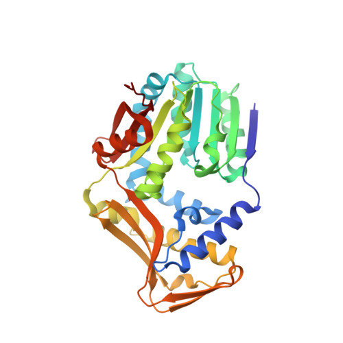
4UY7

Crystal structure of Histidine bound Histidine-specific methyltransferase EgtD from Mycobacterium smegmatis


Experimental Data Snapshot

  • Method: X-RAY DIFFRACTION
  • Resolution: 2.31 Å
  • R-Value Free: 0.247 
  • R-Value Work: 0.192 
  • R-Value Observed: 0.196 

Starting Model: experimental
View more details

wwPDB Validation   3D Report Full Report


Ligand Structure Quality Assessment 


This is version 1.3 of the entry. See complete history


Literature

Structural Insights Into the Histidine Trimethylation Activity of Egtd from Mycobacterium Smegmatis.

Jeong, J.H.Cha, H.J.Ha, S.C.Rojviriya, C.Kim, Y.G.

(2014) Biochem Biophys Res Commun 452: 1098

  • DOI: https://doi.org/10.1016/j.bbrc.2014.09.058
  • Primary Citation of Related Structures:  
    4UY5, 4UY6, 4UY7

  • PubMed Abstract: 

    EgtD is an S-adenosyl-l-methionine (SAM)-dependent histidine N,N,N-methyltransferase that catalyzes the formation of hercynine from histidine in the ergothioneine biosynthetic process of Mycobacterium smegmatis. Ergothioneine is a secreted antioxidant that protects mycobacterium from oxidative stress. Here, we present three crystal structures of EgtD in the apo form, the histidine-bound form, and the S-adenosyl-l-homocysteine (SAH)/histidine-bound form. The study revealed that EgtD consists of two distinct domains: a typical methyltransferase domain and a unique substrate binding domain. The histidine binding pocket of the substrate binding domain primarily recognizes the imidazole ring and carboxylate group of histidine rather than the amino group, explaining the high selectivity for histidine and/or (mono-, di-) methylated histidine as substrates. In addition, SAM binding to the MTase domain induced a conformational change in EgtD to facilitate the methyl transfer reaction. The structural analysis provides insights into the putative catalytic mechanism of EgtD in a processive trimethylation reaction.


  • Organizational Affiliation

    Pohang Accelerator Laboratory, Pohang University of Science and Technology, Pohang, Kyungbuk 790-784, Republic of Korea.


Macromolecules
Find similar proteins by:  (by identity cutoff)  |  3D Structure
Entity ID: 1
MoleculeChains Sequence LengthOrganismDetailsImage
HISTIDINE-SPECIFIC METHYLTRANSFERASE EGTD
A, B
328Mycolicibacterium smegmatisMutation(s): 0 
EC: 2.1.1.44
UniProt
Find proteins for A0R5M8 (Mycolicibacterium smegmatis (strain ATCC 700084 / mc(2)155))
Explore A0R5M8 
Go to UniProtKB:  A0R5M8
Entity Groups  
Sequence Clusters30% Identity50% Identity70% Identity90% Identity95% Identity100% Identity
UniProt GroupA0R5M8
Sequence Annotations
Expand
  • Reference Sequence
Experimental Data & Validation

Experimental Data

  • Method: X-RAY DIFFRACTION
  • Resolution: 2.31 Å
  • R-Value Free: 0.247 
  • R-Value Work: 0.192 
  • R-Value Observed: 0.196 
  • Space Group: P 41
Unit Cell:
Length ( Å )Angle ( ˚ )
a = 74.499α = 90
b = 74.499β = 90
c = 138.954γ = 90
Software Package:
Software NamePurpose
PHENIXrefinement
HKL-2000data reduction
SCALEPACKdata scaling
PHENIXphasing

Structure Validation

View Full Validation Report


Ligand Structure Quality Assessment 


Entry History 

Deposition Data

Revision History  (Full details and data files)

  • Version 1.0: 2014-10-08
    Type: Initial release
  • Version 1.1: 2014-10-15
    Changes: Database references
  • Version 1.2: 2017-07-12
    Changes: Refinement description
  • Version 1.3: 2024-01-10
    Changes: Data collection, Database references, Derived calculations, Other, Refinement description