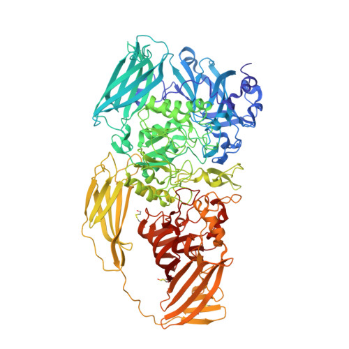
4V41

E. COLI (LAC Z) BETA-GALACTOSIDASE (NCS CONSTRAINED MONOMER-MONOCLINIC)


Experimental Data Snapshot

  • Method: X-RAY DIFFRACTION
  • Resolution: 2.50 Å
  • R-Value Free: 0.207 
  • R-Value Work: 0.199 

wwPDB Validation   3D Report Full Report


This is version 1.3 of the entry. See complete history


Literature

High resolution refinement of beta-galactosidase in a new crystal form reveals multiple metal-binding sites and provides a structural basis for alpha-complementation.

Juers, D.H.Jacobson, R.H.Wigley, D.Zhang, X.J.Huber, R.E.Tronrud, D.E.Matthews, B.W.

(2000) Protein Sci 9: 1685-1699

  • DOI: https://doi.org/10.1110/ps.9.9.1685
  • Primary Citation of Related Structures:  
    1DP0, 1F4A, 1F4H, 4V41

  • PubMed Abstract: 

    The unrefined fold of Escherichia coli beta-galactosidase based on a monoclinic crystal form with four independent tetramers has been reported previously. Here, we describe a new, orthorhombic form with one tetramer per asymmetric unit that has permitted refinement of the structure at 1.7 A resolution. This high-resolution analysis has confirmed the original description of the structure and revealed new details. An essential magnesium ion, identified at the active site in the monoclinic crystals, is also seen in the orthorhombic form. Additional putative magnesium binding sites are also seen. Sodium ions are also known to affect catalysis, and five putative binding sites have been identified, one close to the active site. In a crevice on the protein surface, five linked five-membered solvent rings form a partial clathrate-like structure. Some other unusual aspects of the structure include seven apparent cis-peptide bonds, four of which are proline, and several internal salt-bridge networks. Deep solvent-filled channels and tunnels extend across the surface of the molecule and pass through the center of the tetramer. Because of these departures from a compact globular shape, the molecule is not well characterized by prior empirical relationships between the mass and surface area of proteins. The 50 or so residues at the amino terminus have a largely extended conformation and mostly lie across the surface of the protein. At the same time, however, segment 13-21 contributes to a subunit interface, and residues 29-33 pass through a "tunnel" formed by a domain interface. Taken together, the overall arrangement provides a structural basis for the phenomenon of alpha-complementation.


  • Organizational Affiliation

    Institute of Molecular Biology, and Department of Physics, University of Oregon, Eugene 97403-1229, USA.


Macromolecules
Find similar proteins by:  (by identity cutoff)  |  3D Structure
Entity ID: 1
MoleculeChains Sequence LengthOrganismDetailsImage
BETA-GALACTOSIDASE
A, B, C, D, E
A, B, C, D, E, F, G, H, I, J, K, L, M, N, O, P
1,023Escherichia coli BL21Mutation(s): 3 
EC: 3.2.1.23
UniProt
Find proteins for P00722 (Escherichia coli (strain K12))
Explore P00722 
Go to UniProtKB:  P00722
Entity Groups  
Sequence Clusters30% Identity50% Identity70% Identity90% Identity95% Identity100% Identity
UniProt GroupP00722
Sequence Annotations
Expand
  • Reference Sequence
Small Molecules
Ligands 1 Unique
IDChains Name / Formula / InChI Key2D Diagram3D Interactions
MG
Query on MG

Download Ideal Coordinates CCD File 
AA [auth F]
BA [auth F]
CA [auth G]
DA [auth G]
EA [auth H]
AA [auth F],
BA [auth F],
CA [auth G],
DA [auth G],
EA [auth H],
FA [auth H],
GA [auth I],
HA [auth I],
IA [auth J],
JA [auth J],
KA [auth K],
LA [auth K],
MA [auth L],
NA [auth L],
OA [auth M],
PA [auth M],
Q [auth A],
QA [auth N],
R [auth A],
RA [auth N],
S [auth B],
SA [auth O],
T [auth B],
TA [auth O],
U [auth C],
UA [auth P],
V [auth C],
VA [auth P],
W [auth D],
X [auth D],
Y [auth E],
Z [auth E]
MAGNESIUM ION
Mg
JLVVSXFLKOJNIY-UHFFFAOYSA-N
Modified Residues  1 Unique
IDChains TypeFormula2D DiagramParent
CME
Query on CME
A, B, C, D, E
A, B, C, D, E, F, G, H, I, J, K, L, M, N, O, P
L-PEPTIDE LINKINGC5 H11 N O3 S2CYS
Experimental Data & Validation

Experimental Data

  • Method: X-RAY DIFFRACTION
  • Resolution: 2.50 Å
  • R-Value Free: 0.207 
  • R-Value Work: 0.199 
  • Space Group: P 1 21 1
Unit Cell:
Length ( Å )Angle ( ˚ )
a = 107.9α = 90
b = 207.5β = 94.7
c = 509.9γ = 90
Software Package:
Software NamePurpose
WEISdata scaling
WEISdata reduction
TNTrefinement

Structure Validation

View Full Validation Report


Entry History 

Deposition Data

Revision History  (Full details and data files)

  • Version 1.0: 2014-07-09
    Type: Initial release
  • Version 1.1: 2014-12-10
    Changes: Other
  • Version 1.2: 2017-11-22
    Changes: Refinement description
  • Version 1.3: 2018-01-31
    Changes: Experimental preparation