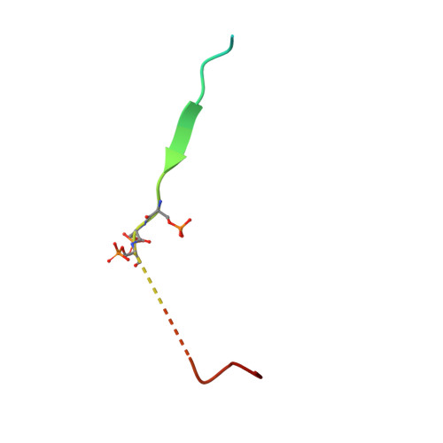
RCSB PDB - 4WJN: Crystal structure of SUMO1 in complex with phosphorylated PML

 4WJN

Crystal structure of SUMO1 in complex with phosphorylated PML


Experimental Data Snapshot

  • Method: X-RAY DIFFRACTION
  • Resolution: 1.50 Å
  • R-Value Free: 0.192 
  • R-Value Work: 0.158 
  • R-Value Observed: 0.160 

Starting Model: experimental
View more details

wwPDB Validation   3D Report Full Report


This is version 1.4 of the entry. See complete history


Literature

Structural and Functional Characterization of the Phosphorylation-Dependent Interaction between PML and SUMO1.

Cappadocia, L.Mascle, X.H.Bourdeau, V.Tremblay-Belzile, S.Chaker-Margot, M.Lussier-Price, M.Wada, J.Sakaguchi, K.Aubry, M.Ferbeyre, G.Omichinski, J.G.

(2015) Structure 23: 126-138

  • DOI: https://doi.org/10.1016/j.str.2014.10.015
  • Primary Citation of Related Structures:  
    4WJN, 4WJO, 4WJP, 4WJQ

  • PubMed Abstract: 

    PML and several other proteins localizing in PML-nuclear bodies (PML-NB) contain phosphoSIMs (SUMO-interacting motifs), and phosphorylation of this motif plays a key role in their interaction with SUMO family proteins. We examined the role that phosphorylation plays in the binding of the phosphoSIMs of PML and Daxx to SUMO1 at the atomic level. The crystal structures of SUMO1 bound to unphosphorylated and tetraphosphorylated PML-SIM peptides indicate that three phosphoserines directly contact specific positively charged residues of SUMO1. Surprisingly, the crystal structure of SUMO1 bound to a diphosphorylated Daxx-SIM peptide indicate that the hydrophobic residues of the phosphoSIM bind in a manner similar to that seen with PML, but important differences are observed when comparing the phosphorylated residues. Together, the results provide an atomic level description of how specific acetylation patterns within different SUMO family proteins can work together with phosphorylation of phosphoSIM's regions of target proteins to regulate binding specificity.


  • Organizational Affiliation

    Département de Biochimie et Médicine Moléculaire, Université de Montréal, C.P. 6128 Succursale Centre-Ville, Montréal, QC H3C 3J7, Canada.


Macromolecules
Find similar proteins by:  (by identity cutoff)  |  3D Structure
Entity ID: 1
MoleculeChains Sequence LengthOrganismDetailsImage
Small ubiquitin-related modifier 183Homo sapiensMutation(s): 1 
Gene Names: SUMO1SMT3CSMT3H3UBL1OK/SW-cl.43
UniProt & NIH Common Fund Data Resources
Find proteins for P63165 (Homo sapiens)
Explore P63165 
Go to UniProtKB:  P63165
PHAROS:  P63165
GTEx:  ENSG00000116030 
Entity Groups  
Sequence Clusters30% Identity50% Identity70% Identity90% Identity95% Identity100% Identity
UniProt GroupP63165
Sequence Annotations
Expand
  • Reference Sequence
Find similar proteins by:  (by identity cutoff)  |  3D Structure
Entity ID: 2
MoleculeChains Sequence LengthOrganismDetailsImage
Protein PML29Homo sapiensMutation(s): 0 
Gene Names: PMLMYLPP8675RNF71TRIM19
EC: 2.3.2
UniProt & NIH Common Fund Data Resources
Find proteins for P29590 (Homo sapiens)
Explore P29590 
Go to UniProtKB:  P29590
PHAROS:  P29590
GTEx:  ENSG00000140464 
Entity Groups  
Sequence Clusters30% Identity50% Identity70% Identity90% Identity95% Identity100% Identity
UniProt GroupP29590
Sequence Annotations
Expand
  • Reference Sequence
Small Molecules
Modified Residues  1 Unique
IDChains TypeFormula2D DiagramParent
SEP
Query on SEP
B
L-PEPTIDE LINKINGC3 H8 N O6 PSER
Experimental Data & Validation

Experimental Data

  • Method: X-RAY DIFFRACTION
  • Resolution: 1.50 Å
  • R-Value Free: 0.192 
  • R-Value Work: 0.158 
  • R-Value Observed: 0.160 
  • Space Group: P 21 21 21
Unit Cell:
Length ( Å )Angle ( ˚ )
a = 38.274α = 90
b = 47.079β = 90
c = 63.986γ = 90
Software Package:
Software NamePurpose
CBASSdata collection
XDSdata scaling
XDSdata reduction
SCALAdata scaling
Cootmodel building
PHENIXphasing
PHENIXrefinement
PDB_EXTRACTdata extraction
XSCALEdata scaling

Structure Validation

View Full Validation Report


Entry History 

Revision History  (Full details and data files)

  • Version 1.0: 2014-12-31
    Type: Initial release
  • Version 1.1: 2015-01-14
    Changes: Database references
  • Version 1.2: 2017-11-22
    Changes: Database references, Derived calculations, Other, Refinement description, Source and taxonomy, Structure summary
  • Version 1.3: 2023-09-27
    Changes: Data collection, Database references, Refinement description
  • Version 1.4: 2024-11-06
    Changes: Structure summary