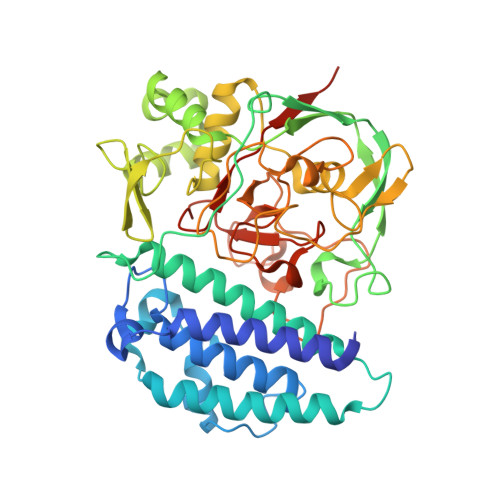
4X8B

Ergothioneine-biosynthetic sulfoxide synthase EgtB, apo form


Experimental Data Snapshot

  • Method: X-RAY DIFFRACTION
  • Resolution: 1.70 Å
  • R-Value Free: 0.157 
  • R-Value Work: 0.138 
  • R-Value Observed: 0.139 

wwPDB Validation   3D Report Full Report


This is version 2.0 of the entry. See complete history


Literature

Structure of the Sulfoxide Synthase EgtB from the Ergothioneine Biosynthetic Pathway.

Goncharenko, K.V.Vit, A.Blankenfeldt, W.Seebeck, F.P.

(2015) Angew Chem Int Ed Engl 54: 2821-2824

  • DOI: https://doi.org/10.1002/anie.201410045
  • Primary Citation of Related Structures:  
    4X8B, 4X8D, 4X8E

  • PubMed Abstract: 

    The non-heme iron enzyme EgtB catalyzes O2 -dependent C-S bond formation between γ-glutamyl cysteine and N-α-trimethyl histidine as the central step in ergothioneine biosynthesis. Both, the catalytic activity and the architecture of EgtB are distinct from known sulfur transferases or thiol dioxygenases. The crystal structure of EgtB from Mycobacterium thermoresistibile in complex with γ-glutamyl cysteine and N-α-trimethyl histidine reveals that the two substrates and three histidine residues serve as ligands in an octahedral iron binding site. This active site geometry is consistent with a catalytic mechanism in which C-S bond formation is initiated by an iron(III)-complexed thiyl radical attacking the imidazole ring of N-α-trimethyl histidine.


  • Organizational Affiliation

    Department for Chemistry, University of Basel, St. Johanns-Ring 19, 4056 Basel (Switzerland).


Macromolecules
Find similar proteins by:  (by identity cutoff)  |  3D Structure
Entity ID: 1
MoleculeChains Sequence LengthOrganismDetailsImage
Sulfoxide synthase EgtB
A, B
447Mycolicibacterium thermoresistibile ATCC 19527Mutation(s): 0 
Gene Names: KEK_08772
EC: 1.14.99.50
UniProt
Find proteins for G7CFI3 (Mycolicibacterium thermoresistibile (strain ATCC 19527 / DSM 44167 / CIP 105390 / JCM 6362 / NCTC 10409 / 316))
Explore G7CFI3 
Go to UniProtKB:  G7CFI3
Entity Groups  
Sequence Clusters30% Identity50% Identity70% Identity90% Identity95% Identity100% Identity
UniProt GroupG7CFI3
Sequence Annotations
Expand
  • Reference Sequence
Small Molecules
Ligands 5 Unique
IDChains Name / Formula / InChI Key2D Diagram3D Interactions
GOL
Query on GOL

Download Ideal Coordinates CCD File 
C [auth A]
D [auth A]
E [auth A]
F [auth A]
G [auth A]
C [auth A],
D [auth A],
E [auth A],
F [auth A],
G [auth A],
L [auth B],
M [auth B],
N [auth B],
O [auth B],
P [auth B]
GLYCEROL
C3 H8 O3
PEDCQBHIVMGVHV-UHFFFAOYSA-N
FE
Query on FE

Download Ideal Coordinates CCD File 
J [auth A],
S [auth B]
FE (III) ION
Fe
VTLYFUHAOXGGBS-UHFFFAOYSA-N
CA
Query on CA

Download Ideal Coordinates CCD File 
K [auth A],
T [auth B]
CALCIUM ION
Ca
BHPQYMZQTOCNFJ-UHFFFAOYSA-N
CL
Query on CL

Download Ideal Coordinates CCD File 
H [auth A],
Q [auth B]
CHLORIDE ION
Cl
VEXZGXHMUGYJMC-UHFFFAOYSA-M
MG
Query on MG

Download Ideal Coordinates CCD File 
I [auth A],
R [auth B]
MAGNESIUM ION
Mg
JLVVSXFLKOJNIY-UHFFFAOYSA-N
Experimental Data & Validation

Experimental Data

  • Method: X-RAY DIFFRACTION
  • Resolution: 1.70 Å
  • R-Value Free: 0.157 
  • R-Value Work: 0.138 
  • R-Value Observed: 0.139 
  • Space Group: P 43 21 2
Unit Cell:
Length ( Å )Angle ( ˚ )
a = 134.78α = 90
b = 134.78β = 90
c = 141.204γ = 90
Software Package:
Software NamePurpose
Aimlessdata scaling
SHELXDEphasing
PHENIXrefinement
PDB_EXTRACTdata extraction
XDSdata reduction
ARPmodel building
Cootmodel building

Structure Validation

View Full Validation Report


Entry History & Funding Information

Deposition Data


Funding OrganizationLocationGrant Number
Swiss National Science FoundationSwitzerland1147005

Revision History  (Full details and data files)

  • Version 1.0: 2015-01-28
    Type: Initial release
  • Version 1.1: 2015-02-25
    Changes: Database references
  • Version 2.0: 2017-09-06
    Changes: Advisory, Atomic model, Author supporting evidence, Data collection, Derived calculations, Refinement description