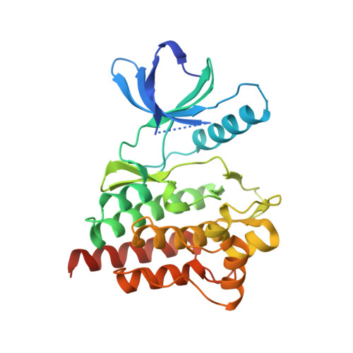
4XG7

Crystal structure of an inhibitor-bound Syk


Experimental Data Snapshot

  • Method: X-RAY DIFFRACTION
  • Resolution: 1.76 Å
  • R-Value Free: 0.242 
  • R-Value Work: 0.223 
  • R-Value Observed: 0.226 

Starting Model: experimental
View more details

wwPDB Validation   3D Report Full Report


Ligand Structure Quality Assessment 


This is version 1.4 of the entry. See complete history


Literature

Crystal structures of spleen tyrosine kinase in complex with novel inhibitors: structural insights for design of anticancer drugs

Lee, S.J.Choi, J.S.Han, B.G.Kim, H.S.Song, H.J.Lee, J.Nam, S.Goh, S.H.Kim, J.H.Koh, J.S.Lee, B.I.

(2016) FEBS J 283: 3613-3625

  • DOI: https://doi.org/10.1111/febs.13831
  • Primary Citation of Related Structures:  
    4XG2, 4XG3, 4XG4, 4XG6, 4XG7, 4XG8, 4XG9, 5GHV

  • PubMed Abstract: 

    Spleen tyrosine kinase (SYK) is a cytosolic nonreceptor protein tyrosine kinase that mediates key signal transduction pathways following the activation of immune cell receptors. SYK regulates cellular events induced by the B-cell receptor and Fc receptors with high intrinsic activity. Furthermore, SYK has been regarded as an attractive target for the treatment of autoimmune diseases and cancers. Here, we report the crystal structures of SYK in complex with seven newly developed inhibitors (G206, G207, O178, O194, O259, O272, and O282) to provide structural insights into which substituents of the inhibitors and binding regions of SYK are essential for lead compound optimization. Our kinase inhibitors exhibited high inhibitory activities against SYK, with half-maximal inhibitory concentrations (IC 50 ) of approximately 0.7-33 nm, but they showed dissimilar inhibitory activities against KDR, RET, JAK2, JAK3, and FLT3. Among the seven SYK inhibitors, O272 and O282 exhibited highly specific inhibitions against SYK, whereas O194 exhibited strong inhibition of both SYK and FLT3. Three inhibitors (G206, G207, and O178) more efficiently inhibited FLT3 while still substantially inhibiting SYK activity. The binding mode analysis suggested that a highly selective SYK inhibitor can be developed by optimizing the functional groups that facilitate direct interactions with Asn499. The atomic coordinates and structure factors for human SYK are in the Protein Data Bank under accession codes 4XG2 (inhibitor-free form), 4XG3 (G206), 4XG4 (G207), 5GHV (O178), 4XG6 (O194), 4XG7 (O259), 4XG8 (O272), and 4XG9 (O282).


  • Organizational Affiliation

    Research Institute, National Cancer Center, Goyang, Gyeonggi, Korea.


Macromolecules
Find similar proteins by:  (by identity cutoff)  |  3D Structure
Entity ID: 1
MoleculeChains Sequence LengthOrganismDetailsImage
Tyrosine-protein kinase SYK290Homo sapiensMutation(s): 0 
Gene Names: SYK
EC: 2.7.10.2
UniProt & NIH Common Fund Data Resources
Find proteins for P43405 (Homo sapiens)
Explore P43405 
Go to UniProtKB:  P43405
PHAROS:  P43405
GTEx:  ENSG00000165025 
Entity Groups  
Sequence Clusters30% Identity50% Identity70% Identity90% Identity95% Identity100% Identity
UniProt GroupP43405
Sequence Annotations
Expand
  • Reference Sequence
Small Molecules
Ligands 1 Unique
IDChains Name / Formula / InChI Key2D Diagram3D Interactions
X7G
Query on X7G

Download Ideal Coordinates CCD File 
B [auth A]1-[(3-methyl-1-{2-[(1-methyl-1H-indazol-5-yl)amino]pyrimidin-4-yl}-1H-pyrazol-4-yl)methyl]azetidin-3-ol
C20 H22 N8 O
FFTGDASJRVPWKO-UHFFFAOYSA-N
Binding Affinity Annotations 
IDSourceBinding Affinity
X7G BindingDB:  4XG7 IC50: min: 15.2, max: 20 (nM) from 2 assay(s)
Experimental Data & Validation

Experimental Data

  • Method: X-RAY DIFFRACTION
  • Resolution: 1.76 Å
  • R-Value Free: 0.242 
  • R-Value Work: 0.223 
  • R-Value Observed: 0.226 
  • Space Group: P 1 21 1
Unit Cell:
Length ( Å )Angle ( ˚ )
a = 39.901α = 90
b = 88.187β = 91.12
c = 40.982γ = 90
Software Package:
Software NamePurpose
REFMACrefinement
HKL-2000data reduction
HKL-2000data scaling
MOLREPphasing

Structure Validation

View Full Validation Report


Ligand Structure Quality Assessment 


Entry History 

Deposition Data

Revision History  (Full details and data files)

  • Version 1.0: 2015-12-30
    Type: Initial release
  • Version 1.1: 2016-08-31
    Changes: Database references
  • Version 1.2: 2016-10-19
    Changes: Database references
  • Version 1.3: 2020-02-05
    Changes: Data collection, Derived calculations
  • Version 1.4: 2023-11-08
    Changes: Data collection, Database references, Refinement description