4XI9 | pdb_00004xi9

Human OGT in complex with UDP-5S-GlcNAc and substrate peptide (RBL2)


Experimental Data Snapshot

  • Method: X-RAY DIFFRACTION
  • Resolution: 3.10 Å
  • R-Value Free: 
    0.223 (Depositor), 0.219 (DCC) 
  • R-Value Work: 
    0.203 (Depositor), 0.199 (DCC) 
  • R-Value Observed: 
    0.203 (Depositor) 

wwPDB Validation 3D Report Full Report

Validation slider image for 4XI9

Ligand Structure Quality Assessment 


This is version 1.3 of the entry. See complete history

Literature

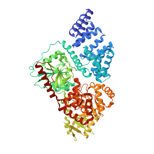
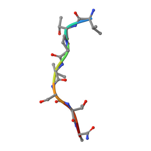
The active site of O-GlcNAc transferase imposes constraints on substrate sequence.

Pathak, S.Alonso, J.Schimpl, M.Rafie, K.Blair, D.E.Borodkin, V.S.Schuttelkopf, A.W.Albarbarawi, O.van Aalten, D.M.

(2015) Nat Struct Mol Biol 22: 744-750

  • DOI: https://doi.org/10.1038/nsmb.3063
  • Primary Citation Related Structures: 
    4XI9, 4XIF, 5BNW, 5C1D

  • PubMed Abstract: 

    O-GlcNAc transferase (OGT) glycosylates a diverse range of intracellular proteins with O-linked N-acetylglucosamine (O-GlcNAc), an essential and dynamic post-translational modification in metazoans. Although this enzyme modifies hundreds of proteins with O-GlcNAc, it is not understood how OGT achieves substrate specificity. In this study, we describe the application of a high-throughput OGT assay to a library of peptides. We mapped sites of O-GlcNAc modification by electron transfer dissociation MS and found that they correlate with previously detected O-GlcNAc sites. Crystal structures of four acceptor peptides in complex with Homo sapiens OGT suggest that a combination of size and conformational restriction defines sequence specificity in the -3 to +2 subsites. This work reveals that although the N-terminal TPR repeats of OGT may have roles in substrate recognition, the sequence restriction imposed by the peptide-binding site makes a substantial contribution to O-GlcNAc site specificity.


  • Organizational Affiliation
    • MRC Protein Phosphorylation and Ubiquitylation Unit and College of Life Sciences, University of Dundee, Dundee, UK.

Macromolecule Content 

  • Total Structure Weight: 329.37 kDa 
  • Atom Count: 22,400 
  • Modeled Residue Count: 2,824 
  • Deposited Residue Count: 2,924 
  • Unique protein chains: 2

Macromolecules

Find similar proteins by:|  3D Structure
Entity ID: 1
MoleculeChains  Sequence LengthOrganismDetailsImage
UDP-N-acetylglucosamine--peptide N-acetylglucosaminyltransferase 110 kDa subunit
A, B, C, D
723Homo sapiensMutation(s): 0 
Gene Names: OGT
EC: 2.4.1.255
UniProt & NIH Common Fund Data Resources
Find proteins for O15294 (Homo sapiens)
Explore O15294 
Go to UniProtKB:  O15294
PHAROS:  O15294
GTEx:  ENSG00000147162 
Entity Groups
Sequence Clusters30% Identity50% Identity70% Identity90% Identity95% Identity100% Identity
UniProt GroupO15294
Sequence Annotations
Expand
Reference Sequence
Find similar proteins by:  Sequence   |   3D Structure  
Entity ID: 2
MoleculeChains  Sequence LengthOrganismDetailsImage
Retinoblastoma-like protein 2
E, F, G, H
8Homo sapiensMutation(s): 0 
UniProt & NIH Common Fund Data Resources
Find proteins for Q08999 (Homo sapiens)
Explore Q08999 
Go to UniProtKB:  Q08999
PHAROS:  Q08999
GTEx:  ENSG00000103479 
Entity Groups
UniProt GroupQ08999
Sequence Annotations
Expand
Reference Sequence

Small Molecules

Ligands 1 Unique
IDChains Name / Formula / InChI Key2D Diagram3D Interactions
12V

Query on 12V


Download:Ideal Coordinates CCD File
I [auth A],
J [auth B],
K [auth C],
L [auth D]
(2S,3R,4R,5S,6R)-3-(acetylamino)-4,5-dihydroxy-6-(hydroxymethyl)tetrahydro-2H-thiopyran-2-yl [(2R,3S,4R,5R)-5-(2,4-dioxo-3,4-dihydropyrimidin-1(2H)-yl)-3,4-dihydroxytetrahydrofuran-2-yl]methyl dihydrogen diphosphate
C17 H27 N3 O16 P2 S
JPRVHSQHWXZSNC-UBDZBXRQSA-N

Experimental Data & Validation

Experimental Data

  • Method: X-RAY DIFFRACTION
  • Resolution: 3.10 Å
  • R-Value Free:  0.223 (Depositor), 0.219 (DCC) 
  • R-Value Work:  0.203 (Depositor), 0.199 (DCC) 
  • R-Value Observed: 0.203 (Depositor) 
Space Group: P 3 2 1
Unit Cell:
Length ( Å )Angle ( ˚ )
a = 273.91α = 90
b = 273.91β = 90
c = 142.93γ = 120
Software Package:
Software NamePurpose
REFMACrefinement
SCALEPACKdata scaling
MOSFLMdata reduction
MOLREPphasing

Structure Validation

View Full Validation Report


Ligand Structure Quality Assessment 


Entry History 

& Funding Information

Deposition Data


Funding OrganizationLocationGrant Number
Wellcome TrustUnited KingdomWT087590MA

Revision History  (Full details and data files)

  • Version 1.0: 2015-08-05
    Type: Initial release
  • Version 1.1: 2015-08-12
    Changes: Database references
  • Version 1.2: 2015-09-16
    Changes: Database references
  • Version 1.3: 2024-05-08
    Changes: Data collection, Database references, Refinement description