4XUF

Crystal structure of the FLT3 kinase domain bound to the inhibitor quizartinib (AC220)


Experimental Data Snapshot

  • Method: X-RAY DIFFRACTION
  • Resolution: 3.20 Å
  • R-Value Free: 0.320 
  • R-Value Work: 0.297 
  • R-Value Observed: 0.299 

wwPDB Validation   3D Report Full Report


Ligand Structure Quality Assessment 


This is version 1.5 of the entry. See complete history


Literature

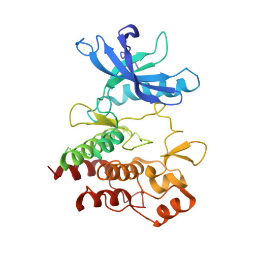
Crystal Structure of the FLT3 Kinase Domain Bound to the Inhibitor Quizartinib (AC220).

Zorn, J.A.Wang, Q.Fujimura, E.Barros, T.Kuriyan, J.

(2015) PLoS One 10: e0121177-e0121177

  • DOI: https://doi.org/10.1371/journal.pone.0121177
  • Primary Citation of Related Structures:  
    4XUF

  • PubMed Abstract: 

    More than 30% of acute myeloid leukemia (AML) patients possess activating mutations in the receptor tyrosine kinase FMS-like tyrosine kinase 3 or FLT3. A small-molecule inhibitor of FLT3 (known as quizartinib or AC220) that is currently in clinical trials appears promising for the treatment of AML. Here, we report the co-crystal structure of the kinase domain of FLT3 in complex with quizartinib. FLT3 with quizartinib bound adopts an "Abl-like" inactive conformation with the activation loop stabilized in the "DFG-out" orientation and folded back onto the kinase domain. This conformation is similar to that observed for the uncomplexed intracellular domain of FLT3 as well as for related receptor tyrosine kinases, except for a localized induced fit in the activation loop. The co-crystal structure reveals the interactions between quizartinib and the active site of FLT3 that are key for achieving its high potency against both wild-type FLT3 as well as a FLT3 variant observed in many AML patients. This co-complex further provides a structural rationale for quizartinib-resistance mutations.


  • Organizational Affiliation

    Department of Molecular and Cell Biology, University of California, Berkeley, California, United States of America; California Institute for Quantitative Biosciences, University of California, Berkeley, California, United States of America.


Macromolecules
Find similar proteins by:  (by identity cutoff)  |  3D Structure
Entity ID: 1
MoleculeChains Sequence LengthOrganismDetailsImage
Receptor-type tyrosine-protein kinase FLT3
A, B
297Homo sapiensMutation(s): 0 
Gene Names: FLT3CD135FLK2STK1
EC: 2.7.10.1
UniProt & NIH Common Fund Data Resources
Find proteins for P36888 (Homo sapiens)
Explore P36888 
Go to UniProtKB:  P36888
PHAROS:  P36888
GTEx:  ENSG00000122025 
Entity Groups  
Sequence Clusters30% Identity50% Identity70% Identity90% Identity95% Identity100% Identity
UniProt GroupP36888
Sequence Annotations
Expand
  • Reference Sequence
Small Molecules
Ligands 1 Unique
IDChains Name / Formula / InChI Key2D Diagram3D Interactions
P30
Query on P30

Download Ideal Coordinates CCD File 
C [auth A],
D [auth B]
1-(5-tert-butyl-1,2-oxazol-3-yl)-3-(4-{7-[2-(morpholin-4-yl)ethoxy]imidazo[2,1-b][1,3]benzothiazol-2-yl}phenyl)urea
C29 H32 N6 O4 S
CVWXJKQAOSCOAB-UHFFFAOYSA-N
Binding Affinity Annotations 
IDSourceBinding Affinity
P30 BindingDB:  4XUF Kd: min: 1.3, max: 420 (nM) from 12 assay(s)
IC50: min: 0.51, max: 1330 (nM) from 24 assay(s)
Experimental Data & Validation

Experimental Data

  • Method: X-RAY DIFFRACTION
  • Resolution: 3.20 Å
  • R-Value Free: 0.320 
  • R-Value Work: 0.297 
  • R-Value Observed: 0.299 
  • Space Group: P 21 21 21
Unit Cell:
Length ( Å )Angle ( ˚ )
a = 48.726α = 90
b = 75.492β = 90
c = 153.968γ = 90
Software Package:
Software NamePurpose
PHENIXrefinement
HKL-2000data reduction
PDB_EXTRACTdata extraction
HKL-2000data scaling
PHENIXphasing

Structure Validation

View Full Validation Report


Ligand Structure Quality Assessment 


Entry History & Funding Information

Deposition Data


Funding OrganizationLocationGrant Number
National Institutes of Health/National Cancer Institute (NIH/NCI)United StatesF32 CA177087-02
Cancer Research InstituteUnited States--
Howard Hughes Medical Institute (HHMI)United States--

Revision History  (Full details and data files)

  • Version 1.0: 2015-04-15
    Type: Initial release
  • Version 1.1: 2015-04-29
    Changes: Database references
  • Version 1.2: 2015-08-26
    Changes: Experimental preparation
  • Version 1.3: 2017-09-06
    Changes: Author supporting evidence, Derived calculations
  • Version 1.4: 2019-11-20
    Changes: Author supporting evidence
  • Version 1.5: 2023-09-27
    Changes: Data collection, Database references, Refinement description, Structure summary