4Y2V | pdb_00004y2v

Structure of soluble epoxide hydrolase in complex with (4-bromo-3-cyclopropyl-1H-pyrazol-1-yl)acetic acid


Experimental Data Snapshot

  • Method: X-RAY DIFFRACTION
  • Resolution: 2.40 Å
  • R-Value Free: 
    0.262 (Depositor), 0.262 (DCC) 
  • R-Value Work: 
    0.200 (Depositor), 0.198 (DCC) 
  • R-Value Observed: 
    0.203 (Depositor) 

Starting Model: experimental
View more details

wwPDB Validation 3D Report Full Report

Validation slider image for 4Y2V

Ligand Structure Quality Assessment 


This is version 1.2 of the entry. See complete history

Literature

Identification of N-ethylmethylamine as a novel scaffold for inhibitors of soluble epoxide hydrolase by crystallographic fragment screening

Amano, Y.Tanabe, E.Yamaguchi, T.

(2015) Bioorg Med Chem 23: 2310-2317

  • DOI: https://doi.org/10.1016/j.bmc.2015.03.083
  • Primary Citation Related Structures: 
    4Y2J, 4Y2P, 4Y2Q, 4Y2R, 4Y2S, 4Y2T, 4Y2U, 4Y2V, 4Y2X, 4Y2Y

  • PubMed Abstract: 

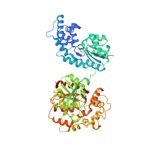
    Soluble epoxide hydrolase (sEH) is a potential target for the treatment of inflammation and hypertension. X-ray crystallographic fragment screening was used to identify fragment hits and their binding modes. Eight fragment hits were identified via soaking of sEH crystals with fragment cocktails, and the co-crystal structures of these hits were determined via individual soaking. Based on the binding mode, N-ethylmethylamine was identified as a promising scaffold that forms hydrogen bonds with the catalytic residues of sEH, Asp335, Tyr383, and Tyr466. Compounds containing this scaffold were selected from an in-house chemical library and assayed. Although the starting fragment had a weak inhibitory activity (IC50: 800μM), we identified potent inhibitors including 2-({[2-(adamantan-1-yl)ethyl]amino}methyl)phenol exhibiting the highest inhibitory activity (IC50: 0.51μM). This corresponded to a more than 1500-fold increase in inhibitory activity compared to the starting fragment. Co-crystal structures of the hit compounds demonstrate that the binding of N-ethylmethylamine to catalytic residues is similar to that of the starting fragment. We therefore consider crystallographic fragment screening to be appropriate for the identification of weak but promising fragment hits.


  • Organizational Affiliation
    • Drug Discovery Research, Astellas Pharma Inc., 21, Miyukigaoka, Tsukuba, Ibaraki 305-8585, Japan. Electronic address: yasushi-amano@astellas.com.

Macromolecule Content 

  • Total Structure Weight: 63.78 kDa 
  • Atom Count: 4,385 
  • Modeled Residue Count: 546 
  • Deposited Residue Count: 561 
  • Unique protein chains: 1

Macromolecules

Find similar proteins by:|  3D Structure
Entity ID: 1
MoleculeChains  Sequence LengthOrganismDetailsImage
Bifunctional epoxide hydrolase 2561Homo sapiensMutation(s): 0 
Gene Names: EPHX2
EC: 3.3.2.10 (PDB Primary Data), 3.1.3.76 (UniProt)
UniProt & NIH Common Fund Data Resources
Find proteins for P34913 (Homo sapiens)
Explore P34913 
Go to UniProtKB:  P34913
PHAROS:  P34913
GTEx:  ENSG00000120915 
Entity Groups
Sequence Clusters30% Identity50% Identity70% Identity90% Identity95% Identity100% Identity
UniProt GroupP34913
Sequence Annotations
Expand
Reference Sequence

Small Molecules

Ligands 2 Unique
IDChains Name / Formula / InChI Key2D Diagram3D Interactions
4A5

Query on 4A5


Download:Ideal Coordinates CCD File
C [auth A](4-bromo-3-cyclopropyl-1H-pyrazol-1-yl)acetic acid
C8 H9 Br N2 O2
DLIOLRMWZAEYPN-UHFFFAOYSA-N
MG

Query on MG


Download:Ideal Coordinates CCD File
B [auth A]MAGNESIUM ION
Mg
JLVVSXFLKOJNIY-UHFFFAOYSA-N
Binding Affinity Annotations 
IDSourceBinding Affinity
4A5 BindingDB:  4Y2V IC50: 9.10e+4 (nM) from 1 assay(s)

Experimental Data & Validation

Experimental Data

  • Method: X-RAY DIFFRACTION
  • Resolution: 2.40 Å
  • R-Value Free:  0.262 (Depositor), 0.262 (DCC) 
  • R-Value Work:  0.200 (Depositor), 0.198 (DCC) 
  • R-Value Observed: 0.203 (Depositor) 
Space Group: P 65 2 2
Unit Cell:
Length ( Å )Angle ( ˚ )
a = 92.203α = 90
b = 92.203β = 90
c = 244.306γ = 120
Software Package:
Software NamePurpose
REFMACrefinement
HKL-2000data reduction
HKL-2000data scaling
AMoREphasing

Structure Validation

View Full Validation Report


Ligand Structure Quality Assessment 


Entry History 

Deposition Data

Revision History  (Full details and data files)

  • Version 1.0: 2015-05-06
    Type: Initial release
  • Version 1.1: 2020-02-05
    Changes: Data collection, Database references, Derived calculations, Source and taxonomy
  • Version 1.2: 2023-11-08
    Changes: Data collection, Database references, Refinement description