4YGJ | pdb_00004ygj

NaBr--Interactions between Hofmeister Anions and the Binding Pocket of a Protein


Experimental Data Snapshot

  • Method: X-RAY DIFFRACTION
  • Resolution: 1.10 Å
  • R-Value Free: 
    0.147 (Depositor), 0.172 (DCC) 
  • R-Value Work: 
    0.127 (Depositor) 
  • R-Value Observed: 
    0.128 (Depositor) 

Starting Model: experimental
View more details

wwPDB Validation 3D Report Full Report

Validation slider image for 4YGJ

This is version 1.5 of the entry. See complete history

Literature

Interactions between Hofmeister Anions and the Binding Pocket of a Protein.

Fox, J.M.Kang, K.Sherman, W.Heroux, A.Sastry, G.M.Baghbanzadeh, M.Lockett, M.R.Whitesides, G.M.

(2015) J Am Chem Soc 137: 3859-3866

  • DOI: https://doi.org/10.1021/jacs.5b00187
  • Primary Citation Related Structures: 
    4YGJ, 4YGK, 4YGL, 4YGN

  • PubMed Abstract: 

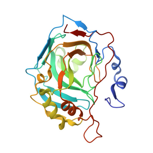
    This paper uses the binding pocket of human carbonic anhydrase II (HCAII, EC 4.2.1.1) as a tool to examine the properties of Hofmeister anions that determine (i) where, and how strongly, they associate with concavities on the surfaces of proteins and (ii) how, upon binding, they alter the structure of water within those concavities. Results from X-ray crystallography and isothermal titration calorimetry show that most anions associate with the binding pocket of HCAII by forming inner-sphere ion pairs with the Zn(2+) cofactor. In these ion pairs, the free energy of anion-Zn(2+) association is inversely proportional to the free energetic cost of anion dehydration; this relationship is consistent with the mechanism of ion pair formation suggested by the "law of matching water affinities". Iodide and bromide anions also associate with a hydrophobic declivity in the wall of the binding pocket. Molecular dynamics simulations suggest that anions, upon associating with Zn(2+), trigger rearrangements of water that extend up to 8 Å away from their surfaces. These findings expand the range of interactions previously thought to occur between ions and proteins by suggesting that (i) weakly hydrated anions can bind complementarily shaped hydrophobic declivities, and that (ii) ion-induced rearrangements of water within protein concavities can (in contrast with similar rearrangements in bulk water) extend well beyond the first hydration shells of the ions that trigger them. This study paints a picture of Hofmeister anions as a set of structurally varied ligands that differ in size, shape, and affinity for water and, thus, in their ability to bind to—and to alter the charge and hydration structure of—polar, nonpolar, and topographically complex concavities on the surfaces of proteins.


  • Organizational Affiliation
    • †Department of Chemistry and Chemical Biology, Harvard University, 12 Oxford Street, Cambridge, Massachusetts 02138, United States.

Macromolecule Content 

  • Total Structure Weight: 29.86 kDa 
  • Atom Count: 2,400 
  • Modeled Residue Count: 258 
  • Deposited Residue Count: 258 
  • Unique protein chains: 1

Macromolecules

Find similar proteins by:|  3D Structure
Entity ID: 1
MoleculeChains  Sequence LengthOrganismDetailsImage
Carbonic anhydrase 2258Homo sapiensMutation(s): 0 
Gene Names: CA2
EC: 4.2.1.1 (PDB Primary Data), 4.2.1.69 (UniProt)
UniProt & NIH Common Fund Data Resources
Find proteins for P00918 (Homo sapiens)
Explore P00918 
Go to UniProtKB:  P00918
PHAROS:  P00918
GTEx:  ENSG00000104267 
Entity Groups
Sequence Clusters30% Identity50% Identity70% Identity90% Identity95% Identity100% Identity
UniProt GroupP00918
Sequence Annotations
Expand
Reference Sequence

Small Molecules

Ligands 2 Unique
IDChains Name / Formula / InChI Key2D Diagram3D Interactions
BR

Query on BR


Download:Ideal Coordinates CCD File
C [auth A]
D [auth A]
E [auth A]
F [auth A]
G [auth A]
C [auth A],
D [auth A],
E [auth A],
F [auth A],
G [auth A],
H [auth A],
I [auth A],
J [auth A],
K [auth A],
L [auth A]
BROMIDE ION
Br
CPELXLSAUQHCOX-UHFFFAOYSA-M
ZN

Query on ZN


Download:Ideal Coordinates CCD File
B [auth A]ZINC ION
Zn
PTFCDOFLOPIGGS-UHFFFAOYSA-N

Experimental Data & Validation

Experimental Data

  • Method: X-RAY DIFFRACTION
  • Resolution: 1.10 Å
  • R-Value Free:  0.147 (Depositor), 0.172 (DCC) 
  • R-Value Work:  0.127 (Depositor) 
  • R-Value Observed: 0.128 (Depositor) 
Space Group: P 1 21 1
Unit Cell:
Length ( Å )Angle ( ˚ )
a = 42.299α = 90
b = 41.379β = 104.64
c = 72.46γ = 90
Software Package:
Software NamePurpose
REFMACrefinement
HKL-2000data reduction
HKL-2000data scaling

Structure Validation

View Full Validation Report


Entry History 

& Funding Information

Deposition Data


Funding OrganizationLocationGrant Number
National Science Foundation (NSF, United States)United StatesNo. 1152196
National Institutes of Health/National Institute of General Medical Sciences (NIH/NIGMS)United States8P41GM103473-16
Department of Energy (DOE, United States)United StatesFWP BO-70
Department of Energy (DOE, United States)United StatesDE-AC02-98CH10886

Revision History  (Full details and data files)

  • Version 1.0: 2015-03-25
    Type: Initial release
  • Version 1.1: 2015-04-08
    Changes: Database references
  • Version 1.2: 2017-08-09
    Changes: Advisory, Derived calculations, Source and taxonomy
  • Version 1.3: 2017-09-06
    Changes: Author supporting evidence
  • Version 1.4: 2019-11-27
    Changes: Author supporting evidence
  • Version 1.5: 2023-09-27
    Changes: Data collection, Database references, Refinement description